
Science Lab
Science Lab
Willkommen auf dem Wissensportal von Leica Microsystems. Hier finden Sie wissenschaftliches Forschungs- und Lehrmaterial rund um das Thema Mikroskopie. Das Portal unterstützt Anfänger, erfahrene Praktiker und Wissenschaftler gleichermaßen bei ihrer täglichen Arbeit und ihren Experimenten. Erkunden Sie interaktive Tutorials und Anwendungshinweise, entdecken Sie die Grundlagen der Mikroskopie ebenso wie High-End-Technologien. Werden Sie Teil der Science Lab Community und teilen Sie Ihr Fachwissen.
Filter articles
Tags
Beitragstyp
Produkte
Loading...

Formulated Product Characterization with SRS Microscopy
Creams, pastes, gels, emulsions, and tablets are ubiquitous across a wide range of manufacturing sectors from pharmaceuticals and consumer health products to agrochemicals and paint. To improve…
Loading...

Querschnitt-Ionenstrahlfräsen von Batteriekomponenten
Für ein umfassendes Verständnis von Lithiumbatteriesystemen ist eine qualitativ hochwertige Oberflächenpräparation erforderlich, um die innere Struktur und Morphologie zu untersuchen. Aufgrund der…
Loading...

High-resolution 3D Imaging to Investigate Tissue Ageing
Award-winning researcher Dr. Anjali Kusumbe demonstrates age-related changes in vascular microenvironments through single-cell resolution 3D imaging of young and aged organs.
Loading...

How to Successfully Perform Live-Cell CLEM
The Leica Nano workflow provides a streamlined live-cell CLEM solution for getting insight bout structural changes of cellular components over time. Besides the technical handling described in the…
Loading...
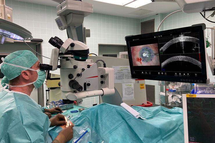
Proveo 8 with intraoperative OCT – a User Evaluation in an University Setting
Optical coherence tomography (OCT) makes structures in the eye visible that lie beneath the surface. When OCT is used intraoperatively, surgeons gain insight into possible pathological changes in the…
Loading...

Be Confident in your Results with Cell DIVE Validated Antibodies
The Cell DIVE System includes a carefully curated list of hundreds of commercially available antibodies validated to offer optimal specificity and sensitivity in multiplexed imaging. That validation…
Loading...
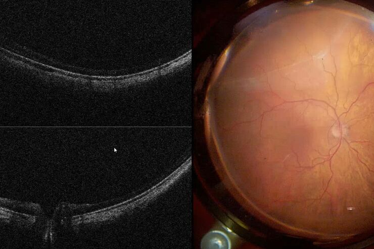
Intraoperative OCT in Retinal Procedures
In their white paper the surgical team at Retina Clinic in São Paulo provide insight into cases of complex retinal surgery in which OCT has provided helpful additional information which has on…
Loading...
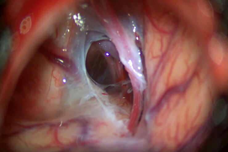
Optimal Visualization in Brain Surgery
This case study “Treatment of the Galassi type III arachnoid cyst with the M530 OHX surgical microscope from Leica Microsystems” documents the procedure step by step and shows the visualization…
Loading...

Benefits of Combining STED and Lifetime
In this interview, Professor Alberto Diaspro talks about the advantages of the White Light Laser and the TauSTED capabilities of STELLARIS 8 STED. He speaks about his experience with the confocal…
