

Science Lab
Science Lab
Willkommen auf dem Wissensportal von Leica Microsystems. Hier finden Sie wissenschaftliches Forschungs- und Lehrmaterial rund um das Thema Mikroskopie. Das Portal unterstützt Anfänger, erfahrene Praktiker und Wissenschaftler gleichermaßen bei ihrer täglichen Arbeit und ihren Experimenten. Erkunden Sie interaktive Tutorials und Anwendungshinweise, entdecken Sie die Grundlagen der Mikroskopie ebenso wie High-End-Technologien. Werden Sie Teil der Science Lab Community und teilen Sie Ihr Fachwissen.
Filter articles
Tags
Beitragstyp
Produkte
Loading...
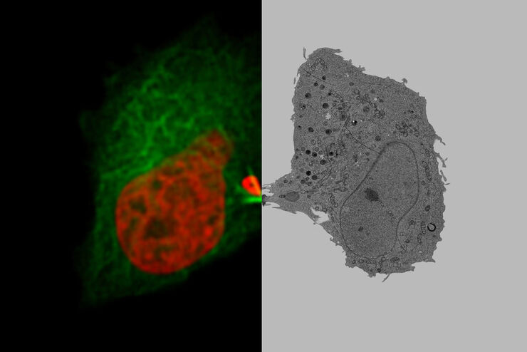
Putting Dynamic Live Cell Data into the Ultrastructural Context
With workflow Coral Life, searching for a needle in the haystack is a thing of the past. Take advantage of correlative light and electron microscopy to identify directly the right cell at the right…
Loading...
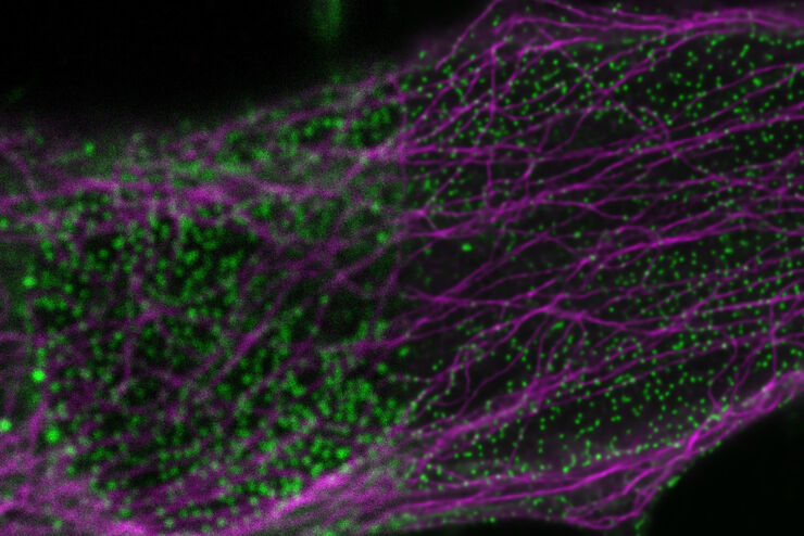
AI in Microscopy Webinar
We demonstrate residual channel attention networks for restoring and enhancing volumetric time-lapse (4D) fluorescence microscopy data.
Loading...

20 Years of Leica Laser Microdissection
Phenotype-genotype correlations are key for insight. From Eye to Insight is therefore fitting perfectly to Leica Microsystems and in particular to laser microdissection. Laser Microdissection, also…
Loading...
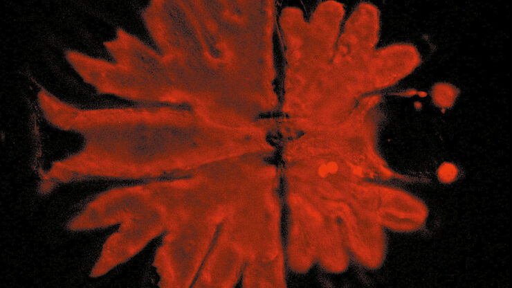
Phasenkontrast
Mit einem Phasenkontrast-Lichtmikroskop können die Strukturen vieler Arten von biologischen Präparaten mit einem größeren Kontrast betrachtet werden, ohne dass die Probe eingefärbt werden muss.
Loading...
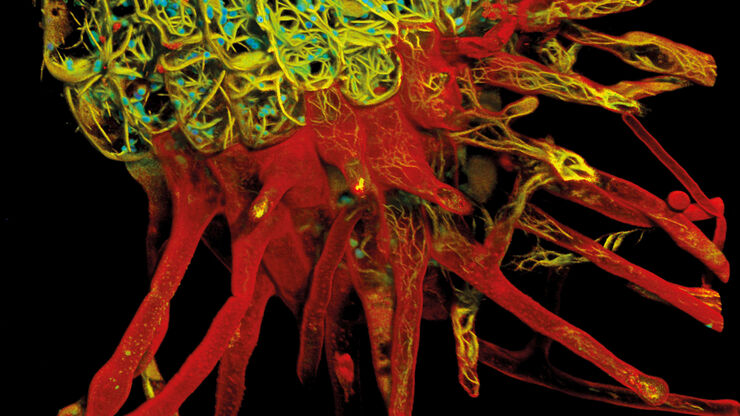
Benefits of TauContrast to Image Complex Samples
In this interview, Dr. Timo Zimmermann talks about his experience with the application of TauSense tools and their potential for the investigation of demanding samples such as thick samples or…
Loading...
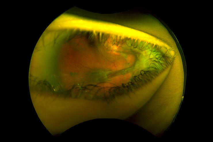
Intraoperative OCT-Assisted Surgical Management of Proliferative Vitreoretinopathy
Proliferative vitreoretinopathy (PVR) is a plague to patients and their surgeons after recent rhegmatogenous retinal detachment (RD). Despite excellent initial surgical outcomes, it is the most common…
Loading...

Fast, High-quality Vitrification with the EM ICE High Pressure Freezer
The EM ICE High Pressure Freezer was developed with a unique freezing principle and uses only a single pressurization and cooling liquid: liquified nitrogen (LN2). This design enables three major…
Loading...
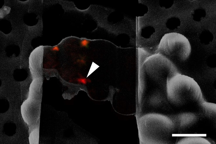
Targeting Active Recycling Nuclear Pore Complexes using Cryo Confocal Microscopy
In this article, how cryo light microscopy and, in particular cryo confocal microscopy, is used to improve the reliability of cryo EM workflows is described. The quality of the EM grids and samples is…
Loading...

How Augmented Reality is Transforming Vascular Neurosurgery
Augmented Reality is changing surgery, with new information helping to improve the precision and safety of procedures. This is especially true in vascular neurosurgery where Augmented Reality is…


