Science Lab

Science Lab

Science Lab

ライカマイクロシステムズのナレッジポータルでは、顕微鏡の基礎から最先端技術まで、幅広い情報を提供しています。初心者から熟練者、研究者、医師の皆様まで、日々の研究や実験に役立つ内容となっております。チュートリアルやアプリケーションノートを活用し、学びながら探究心を刺激してください。さらに、コミュニティに参加することで、知見を共有し、新たな発見へとつなげましょう。お気軽に参加いただき、互いの専門知識を深め合う場としてご活用ください。

From Organs to Tissues to Cells: Analyzing 3D Specimens with Widefield Microscopy

Obtaining high-quality data and images from thick 3D samples is challenging using traditional widefield microscopy because of the contribution of out-of-focus light. In this webinar, Falco Krüger…
Mouse retina was fixed and stained by following reagents: anti-CD31 antibody (green): Endothelia cells, IsoB4 (red): Blood vessels, and microglia anti-GFAP antibody (blue): Astrocytes Sample courtesy by Jeremy Burton, PhD and Jiyeon Lee, PhD, Genentech Inc., South San Francisco, USA. Imaged by Olga Davydenko, PhD (Leica). Imaged with a THUNDER Imager 3D Cell Culture.

An Introduction to Computational Clearing

Many software packages include background subtraction algorithms to enhance the contrast of features in the image by reducing background noise. The most common methods used to remove background noise…

Factors to Consider When Selecting a Research Microscope

An optical microscope is often one of the central devices in a life-science research lab. It can be used for various applications which shed light on many scientific questions. Thereby the…
Influenca in lung epithelial cells (porcine) - THUNDER Imager 3D Cell Culture Influenca virus – red, cilia – green, Nuclei – blue.

How Can Immunofluorescence Aid Virology Research?

Modern virology research has become as crucial now as ever before due to the global COVID-19 pandemic. There are many powerful technologies and assays that virologists can apply to their research into…

Computational Clearing - Enhance 3D Specimen Imaging

This webinar is designed to clarify crucial specifications that contribute to THUNDER Imagers' transformative visualization of 3D samples and improvements within a researcher's imaging-related…

THUNDER Imagers: High Performance, Versatility and Ease-of-Use for your Everyday Imaging Workflows

This webinar will showcase the versatility and performance of THUNDER Imagers in many different life science applications: from counting nuclei in retina sections and RNA molecules in cancer tissue…

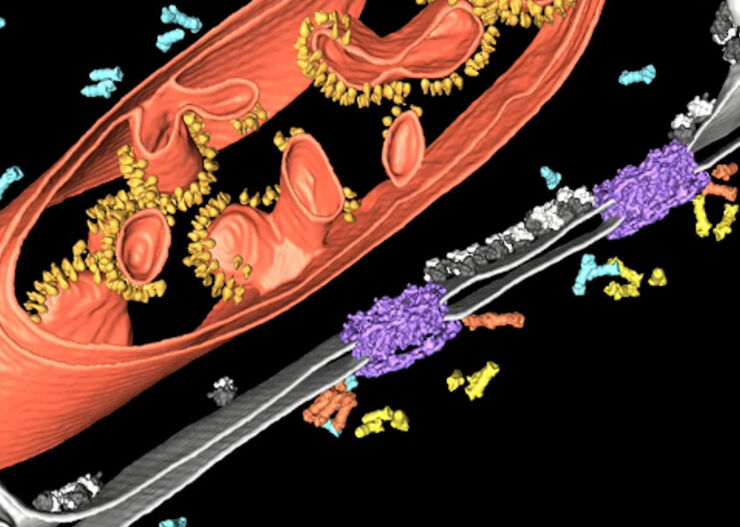
Improve Cryo Electron Tomography Workflow

Leica Microsystems and Thermo Fisher Scientific have collaborated to create a fully integrated cryo-tomography workflow that responds to these research needs: Reveal cellular mechanisms at…

アルツハイマー病プラーク:厚みのある組織切片での高速可視化

認知症と診断された症例の60%以上がアルツハイマー病ということが知られています。この病気の典型は、脳組織の組織学的変化にあります。今のところ、この病気に対する確固たる治療法はありません。今行われている治療法は、致命的な進行を遅らせることや、患者の症状を和らげようとするものです。Mehrdad…
HeLa cell spheroid stained with Alexa Fluor 568 Phalloidin (Actin) and YOYO 1 iodide (Nucleus).

Real Time Images of 3D Specimens with Sharp Contrast Free of Haze

THUNDER Imagers deliver in real time images of 3D specimens with sharp contrast, free of the haze or out-of-focus blur typical of widefield systems. They can even image clearly places deep inside a…
Scroll to top