
Life Science Research
Life Science Research
This is the place to expand your knowledge, research capabilities, and practical applications of microscopy in various scientific fields. Learn how to achieve precise visualization, image interpretation, and research advancements. Find insightful information on advanced microscopy, imaging techniques, sample preparation, and image analysis. Topics covered include cell biology, neuroscience, and cancer research with a focus on cutting-edge applications and innovations.

A Practical Guide to Virology Research
Leica solutions for imaging and sample preparation help you with the investigation of viral entry and fusion, genome integration, viral replication, assembly, and virus budding.

Microscopy in Virology
The coronavirus SARS-CoV-2, causing the Covid-19 disease effects our world in all aspects. Research to find immunization and treatment methods, in other words to fight this virus, gained highest…

Computational Clearing - Enhance 3D Specimen Imaging
This webinar is designed to clarify crucial specifications that contribute to THUNDER Imagers' transformative visualization of 3D samples and improvements within a researcher's imaging-related…
Explore Innovative Techniques to Separate Fluorophores with Overlapping Spectra
In this article we explore several strategies you can take to improve the separation of fluorophores and increase the number of fluorescent probes you can distinguish in your sample.

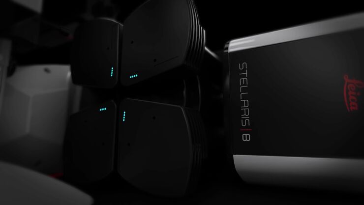
STELLARIS White Light Lasers
When it comes to choosing fluorescent probes for your multi-color experiments, you shouldn’t have to compromise. Now you can advance beyond conventional excitation sources that limit your fluorophore…

TauSense Technology Imaging Tools
Leica Microsystems’ TauSense technology is a set of imaging modes based on fluorescence lifetime. Found at the core of the STELLARIS confocal platform, it will revolutionize your imaging experiments.…
The Power HyD Detector Family
Powerful photon counting detectors on the STELLARIS confocal platform provide improved photon counting, ultra-sensitive imaging and more color options in the NIR spectrum.
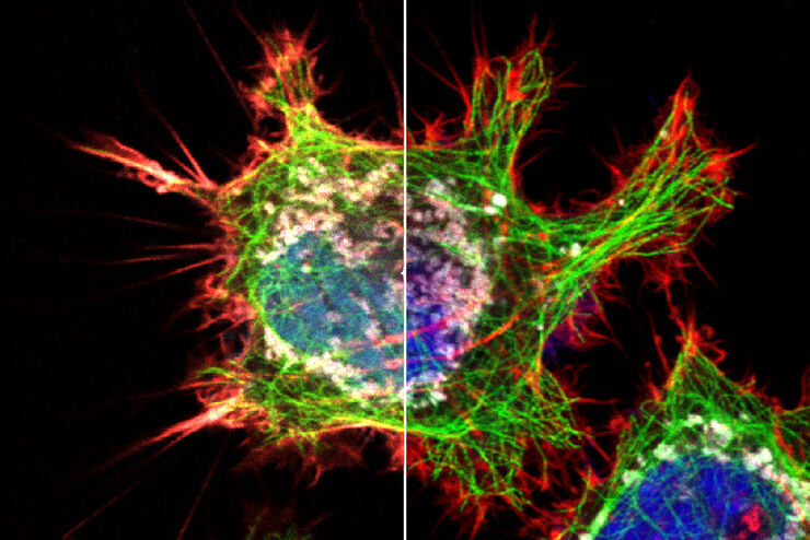
, and trafficking vesicles labeled with CF594 (cyan - Biotium).")
A Guide to Super-Resolution
Find out more about Leica super-resolution microscopy solutions and how they can empower you to visualize in fine detail subcellular structures and dynamics.

Guide to Microscopy in Cancer Research
Cancer is a complex and heterogeneous disease caused by cells deficient in growth regulation. Genetic and epigenetic changes in one or a group of cells disrupt normal function and result in…
