Life Science Research
Life Science Research
This is the place to expand your knowledge, research capabilities, and practical applications of microscopy in various scientific fields. Learn how to achieve precise visualization, image interpretation, and research advancements. Find insightful information on advanced microscopy, imaging techniques, sample preparation, and image analysis. Topics covered include cell biology, neuroscience, and cancer research with a focus on cutting-edge applications and innovations.
A Guide to Using Microscopy for Drosophila (Fruit Fly) Research
The fruit fly, typically Drosophila melanogaster, has been used as a model organism for over a century. One reason is that many disease-related genes are shared between Drosophila and humans. It is…
Selecting the Right Dissecting Microscope
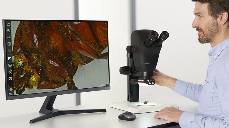
Learn how you can enhance dissection for life-science research and education with a microscope that ensures ergonomic comfort, high-quality optics, and easy access to the specimen.
Depth of Field in Microscope Images
For microscopy imaging, depth of field is an important parameter when needing sharp images of sample areas with structures having significant changes in depth. In practice, depth of field is…
Understanding Clearly the Magnification of Microscopy
To help users better understand the magnification of microscopy and how to determine the useful range of magnification values for digital microscopes, this article provides helpful guidelines.
Key Factors to Consider When Selecting a Stereo Microscope
This article explains key factors that help users determine which stereo microscope solution can best meet their needs, depending on the application.
What is the FusionOptics Technology?
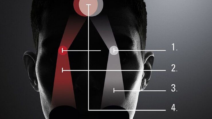
Leica stereo microscopes with FusionOptics provide optimal 3D perception. The brain merges two images, one with large depth of field and the other with high resolution, into one 3D image.
Microscope Ergonomics
This article explains microscope ergonomics and how it helps users work in comfort, enabling consistency and efficiency. Learn how to set up the workplace to keep good posture when using a microscope.
Rodent and Small-Animal Surgery
Learn how you can perform rodent (mouse, rat, hamster) and small-animal surgery efficiently with a microscope for developmental biology and medical research applications by reading this article.
Factors for Selecting Student Microscopes
If chosen carefully, educational microscopes will open windows to a cosmos of minute detail which delight young minds in schools and universities – and ideally keeps them fascinated enough to make…

