
Filter articles
タグ
製品
Loading...
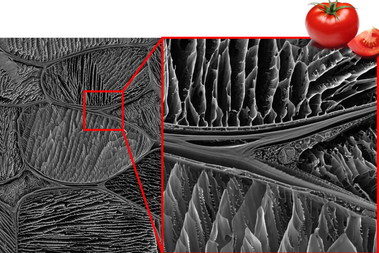
Studying the Microstructure of Natural Polymers in Fine Detail
The potential of cryogenic broad ion beam milling used in combination with scanning electron microscopy (cryo-BIB-SEM) for imaging and analyzing the microstructure of cryogenically stabilized soft…
Loading...
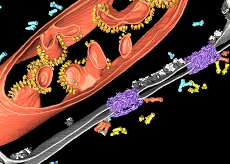
Improve Cryo Electron Tomography Workflow
Leica Microsystems and Thermo Fisher Scientific have collaborated to create a fully integrated cryo-tomography workflow that responds to these research needs: Reveal cellular mechanisms at…
Loading...
![3D glomeruli in a portion of an ECi-cleared kidney scanned by light sheet microscopy. Courtesy of Prof. Norbert Gretz, Medical Faculty Mannheim, University of Heidelberg [1].](/fileadmin/_processed_/d/d/csm_DLS-Sample-Preparation-Intr_915e0fd7c2.jpg "3D glomeruli in a portion of an ECi-cleared kidney scanned by light sheet microscopy. Courtesy of Prof. Norbert Gretz, Medical Faculty Mannheim, University of Heidelberg [1].")
Using Mounting Frames for Light Sheet Sample Preparation
Sample handling is an important topic in the context of Light Sheet Microscopy. The TCS SP8 DLS integrates Light Sheet technology into an inverted confocal platform and can hence make use of general…
Loading...

Using a Rotation Device for Light Sheet Sample Mounting
The TCS SP8 DLS from Leica Microsystems is an innovative concept to integrate the Light Sheet Microscopy technology into the confocal microscope. Due to its unique optical architecture samples can be…
Loading...

High Resolution Array Tomography with Automated Serial Sectioning
The optimization of high resolution, 3-dimensional (3D), sub-cellular structure analysis with array tomography using an automated serial sectioning solution, achieving a high section density on the…
Loading...

Macroscale to Nanoscale Pore Analysis of Shale and Carbonate Rocks
Physical porosity in rocks, like shale and carbonate, has a large effect on the their storage capacity. The pore geometries also affect their permeability. Imaging the visible pore space provides…
Loading...

Using U-Shaped Glass Capillaries for Sample Mounting
The DLS microscope system from Leica Microsystems is an innovative concept which integrates the Light Sheet Microscopy technology into the confocal platform. Due to its unique optical architecture,…
Loading...
Each Atom Counts: Protect Your Samples Prior to FIB Processing
Application Note for Leica EM ACE600 - Focused ion beam (FIB) technology has become an indispensable tool for site-specific TEM sample preparation. It allows to extract electron transparent specimens…
Loading...
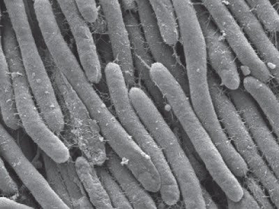
Bacteria Protocol - Critical Point Drying of E. coli for SEM
Application Note for Leica EM CPD300 - Critical point drying of E. coli with subsequent platinum / palladium coating and SEM analysis. Sample was inserted into a filter disc (Pore size: 16 - 40 μm)…
