Compound Light Microscopes
Compound Light Microscopes
What are you interested in? Show subnavigation
Compound Light Microscopes


Products Show subnavigation
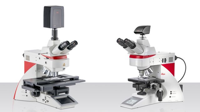
DM4 B & DM6 B
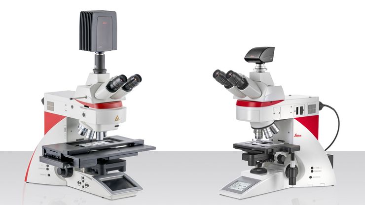
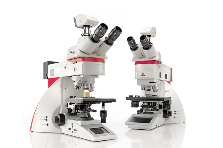
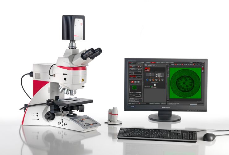
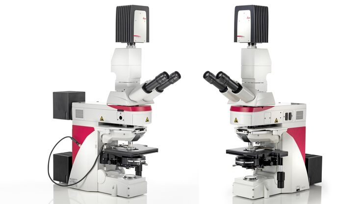
Enhance your life-science and clinical research with the DM4 B and DM6 B upright microscopes. With flexible software solutions, they allow you to focus on experiments while reducing manual work.
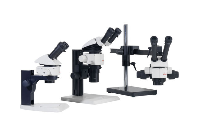
M50, M60 & M80
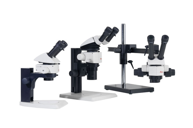
Routine stereo microscopes
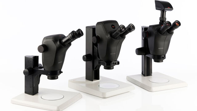
Ivesta 3
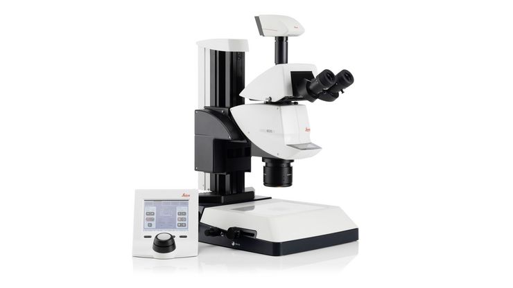
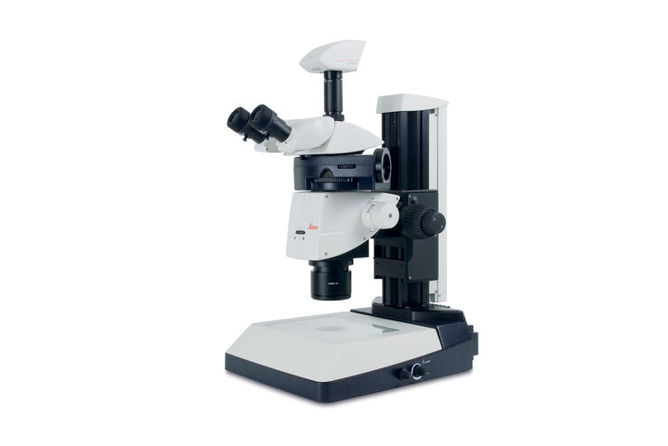
Greenough Stereo Microscopes. Optimize visual inspection and rework while achieving reliable, consistent results.

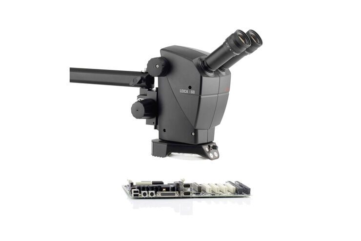
A60 F & A60 S
Industrial Stereo Microscopes for Electronics and Medical Device Manufacturing
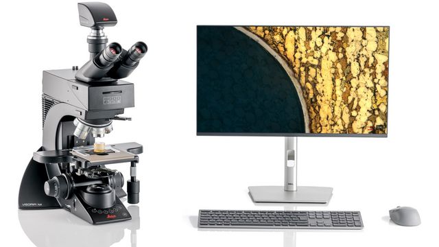
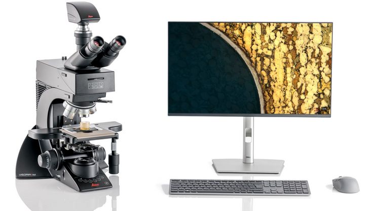
Visoria M
Experience enhanced efficiency and comfort in your daily microscopy routine. The Visoria M materials microscope is for applications performed in the metal, electronics, and polymer industries as well...

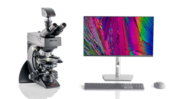
Visoria P
Experience enhanced efficiency and comfort in your daily microscopy routine. The Visoria P polarization microscope uses polarized light to study the optical properties of materials and geological...
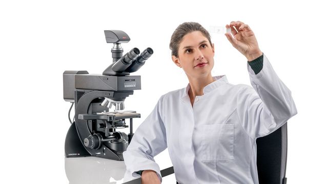
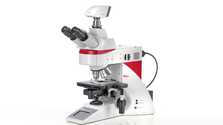
Visoria B
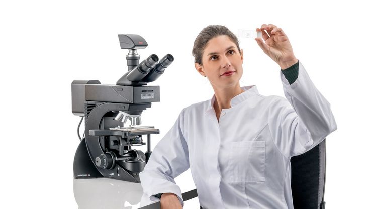
Experience enhanced efficiency and comfort in your daily microscopy routine. The Visoria B laboratory microscope is for applications performed in life science and clinical laboratories.

THUNDER Imager Cell Spinning Disk
Discover the power of combining THUNDER technology with the CrestOptics CICERO spinning disk.
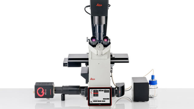
DMi8
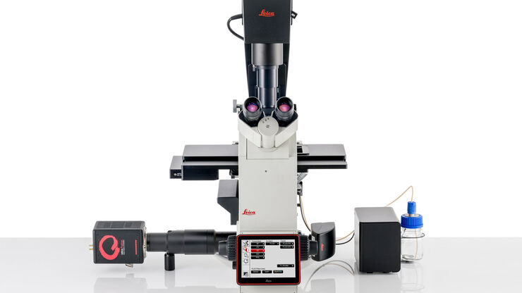
DMi8 inverted microscope platform enables you to generate high quality data with solutions tailored to your research requirements and budget.
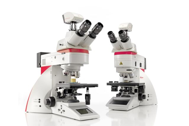
Leica DM4 M & DM6 M
Upright Materials Microscopes
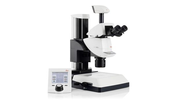
M125 C, M205 C & M205 A
Encoded Stereo Microscopes

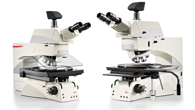
DM8000 M & DM12000 M
Detect defects and carry out sample overviews quickly with the reliable DM8000 M & DM12000 M inspection microscopes.
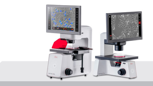
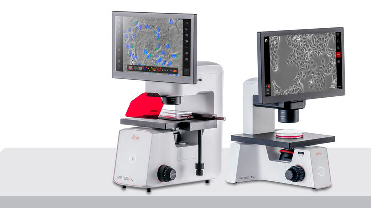
Mateo Fl, Mateo TL
Integrated inverted digital microscopes for cell culture research with AI-assisted workflows for consistent cell culture analysis.
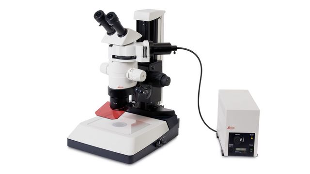
MZ10 F
Modular stereomicroscope for fluorescent imaging
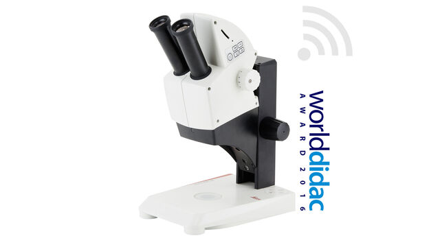
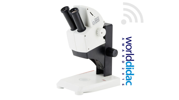
Leica EZ4 W & EZ4 E
Stereo microscope for college and university education with 8x to 35x magnification, 7-way LED illumination, and integrated camera
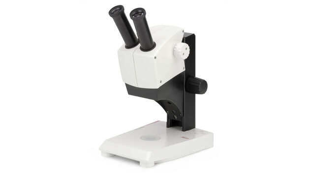
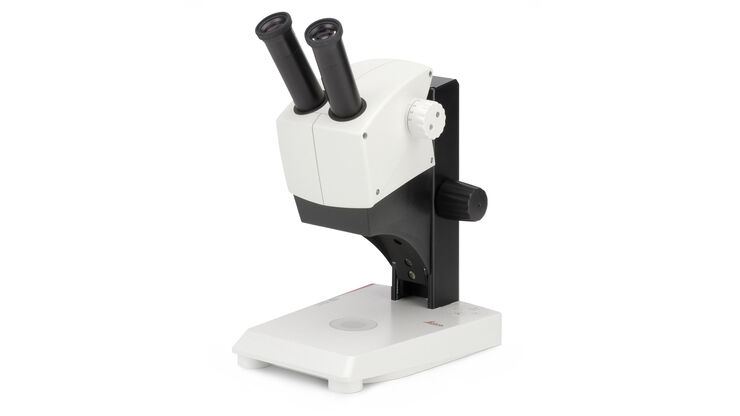
EZ4
Stereo microscope for beginners in college and university education with 8x to 35x magnification and 7-way LED illumination
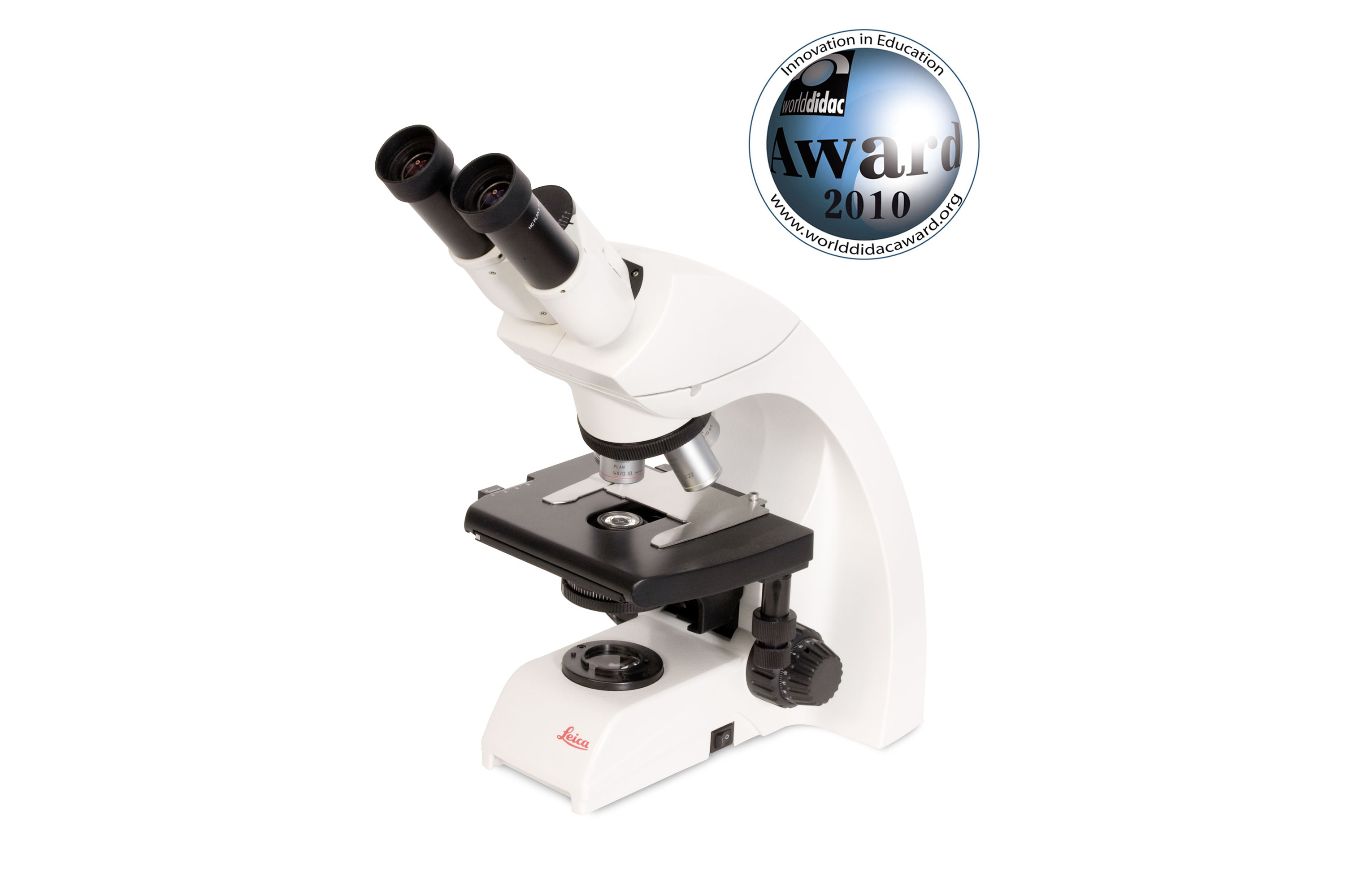
DM500
Binocular educational microscope for life science students with 4 infinity-corrected plan-achromat objectives and fluorescence capability
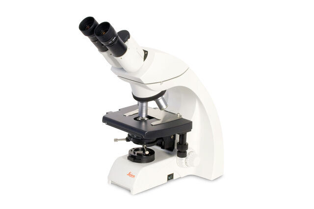
DM750
Binocular educational microscope for life science postdocs with 4 or 5 infinity-corrected or HI plan-achromat objectives and fluorescence capability
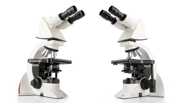
DM1000
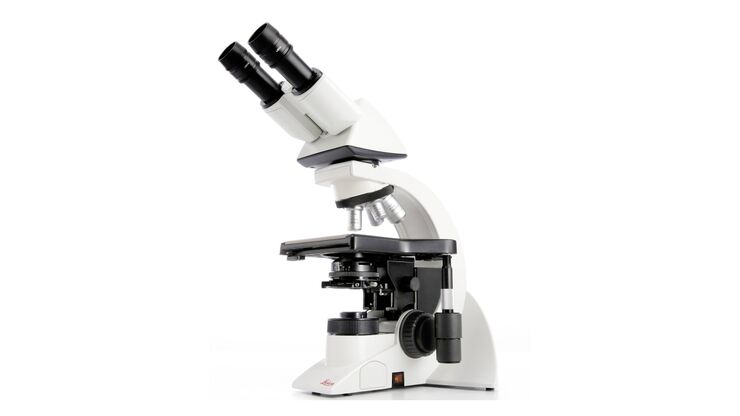
Uniquely Ergonomic System Microscope
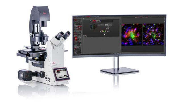
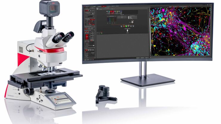
THUNDER Imager Cell
THUNDER Imagers provide you with a solution for advanced 3D cell culture assays, whether you want to study stem cells, spheroids, or organoids.
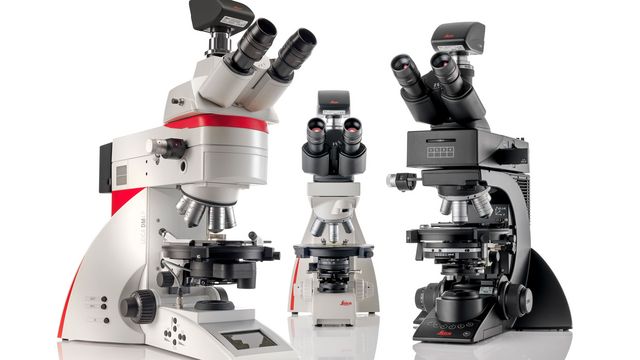
DM4 P, DM750 P & Visoria P
Polarization Microscopes
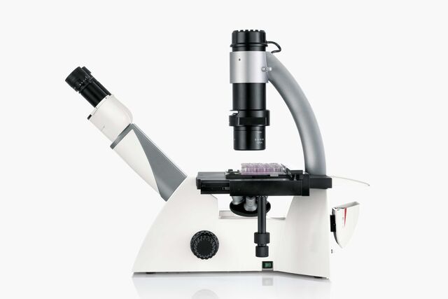
DMi1
Entry level inverted microscope
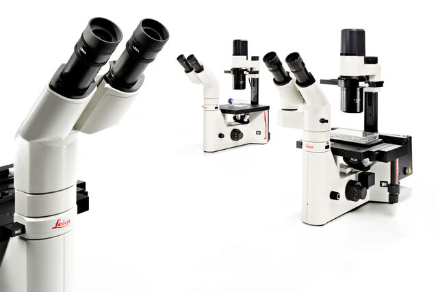
DM IL LED
Inverted Laboratory Microscope with LED Illumination
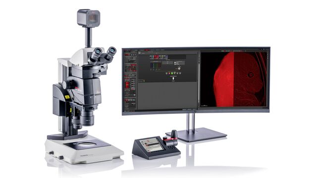
THUNDER Imager Model Organism
The THUNDER Imager Model Organism allows fast and easy 3D exploration of whole organisms for developmental or molecular biology research.
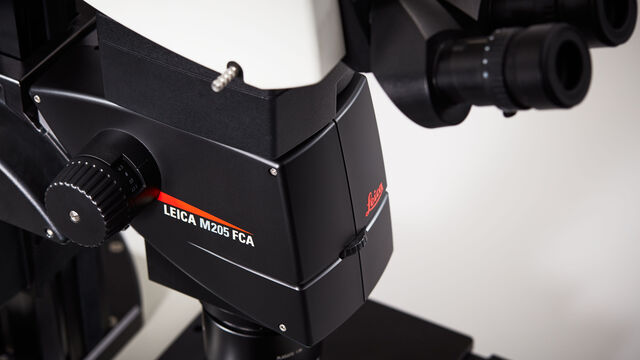
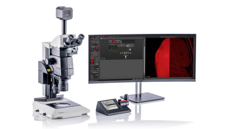
Leica M205 FCA & Leica M205 FA
(Semi)-automated fluorescence stereo microscopes
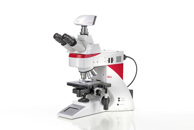
Leica DM4 B & DM6 B
Increase work efficiency with Leica DM4 B & DM6 B upright digital research microscopes.
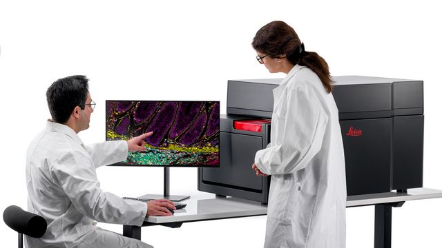
Cell DIVE
Multiplex imaging solution Cell DIVE offers crystal-clear whole tissue images, the visualization of 60+ biomarkers and over 350 validated antibodies.
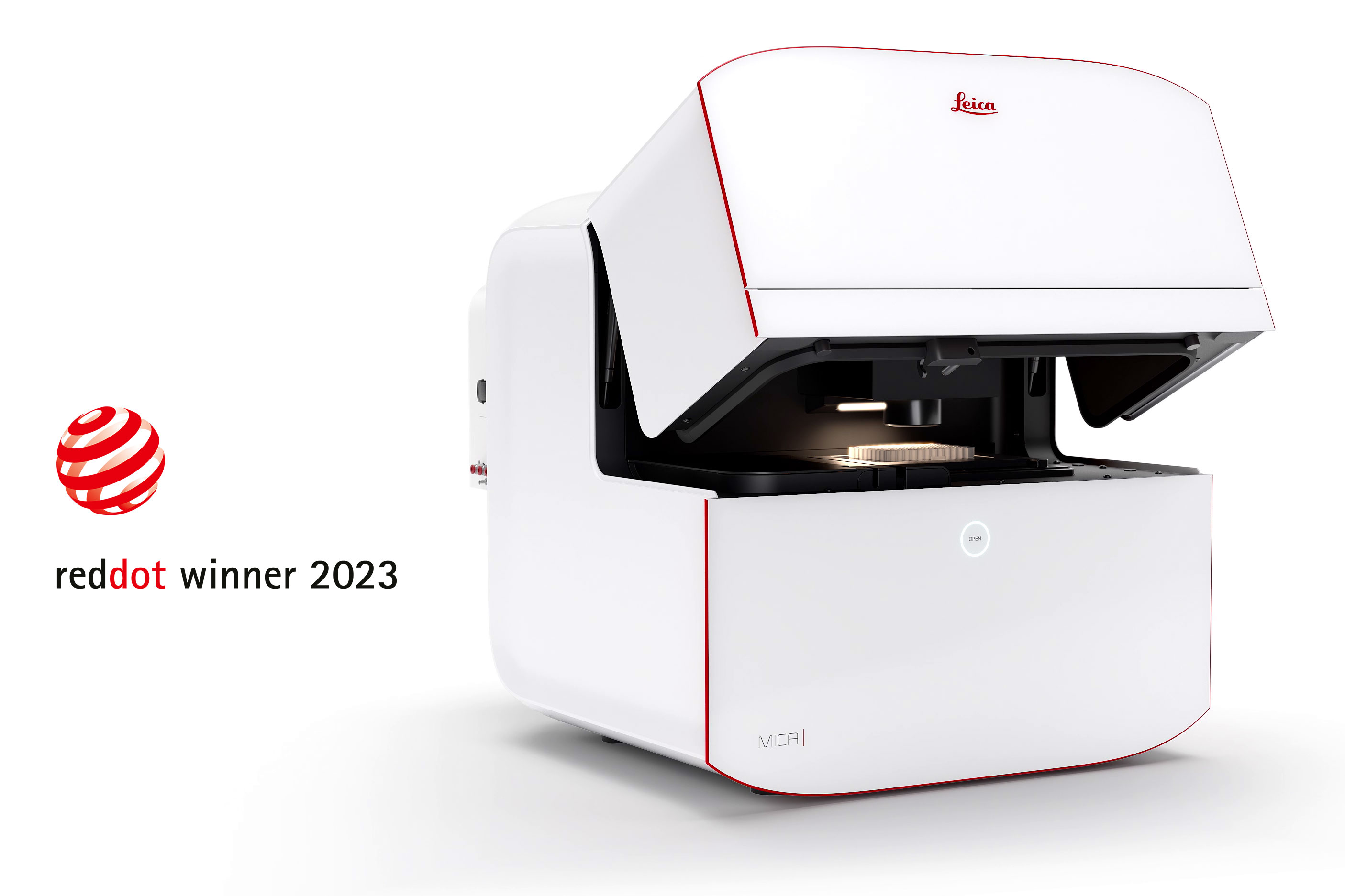
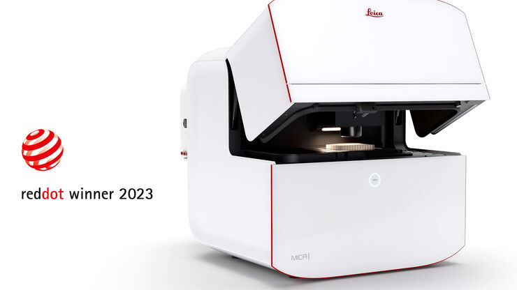
Mica
Mica - The world’s first Microhub. Everything you need to enable discoveries, unified in one easy-to-use system. Simultaneous 4-color widefield, confocal resolution, AI supported analysis.
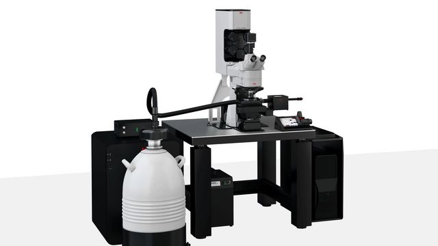
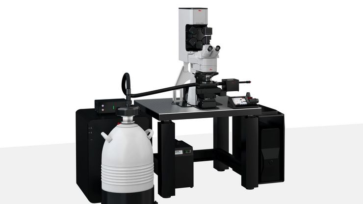
STELLARIS Cryo
STELLARIS Cryo is a confocal light microscope system that helps you to target your area of interest for cryo-electron tomography (CryoET)
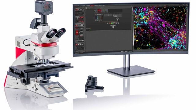
THUNDER Imager Tissue
The THUNDER Imager Tissue allows real-time fluorescence imaging of 3D tissue sections typically used in neuroscience and histology research.
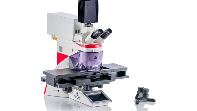
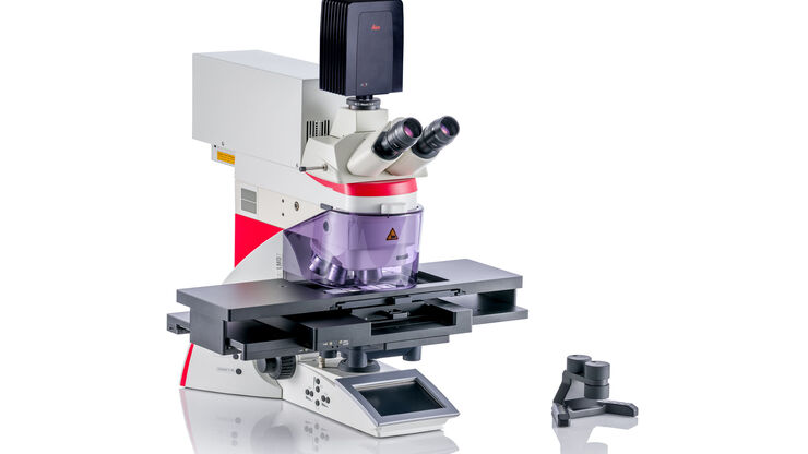
Leica LMD Systems
Laser Microdissection enables users to isolate specific single cells or entire areas of tissue.
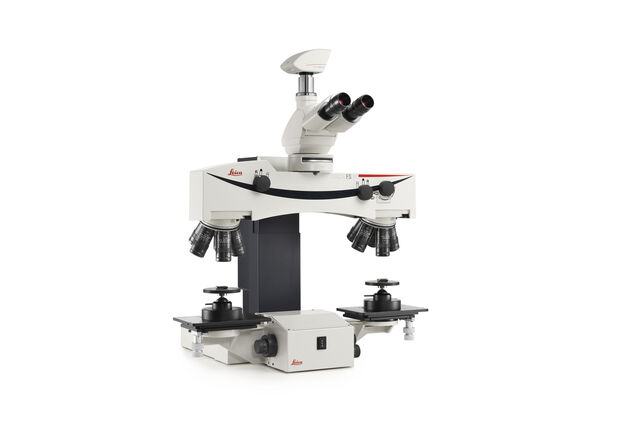
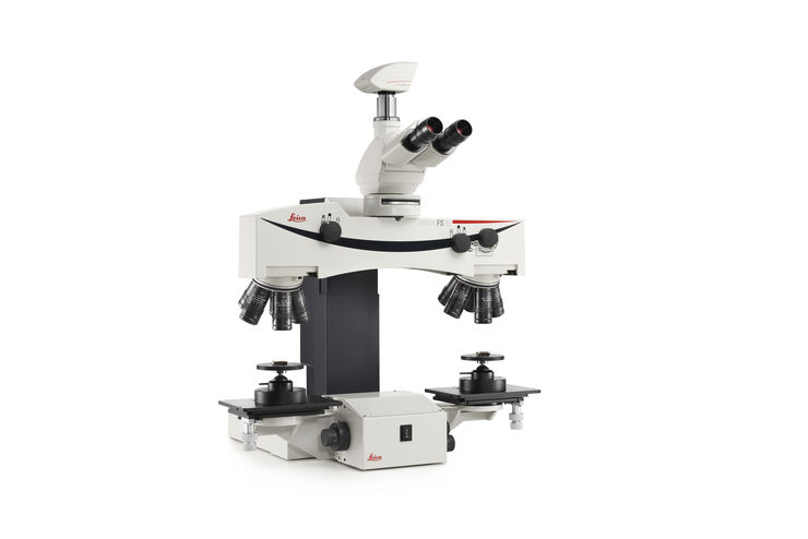
FS M
Manual Forensic Comparison Macroscope

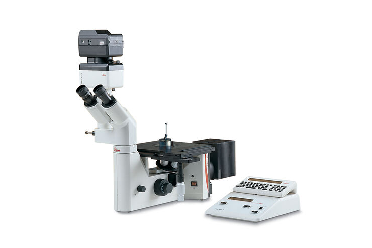
DM ILM
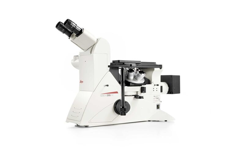
Inverted Microscope for Metallography and Industrial Materials Inspection
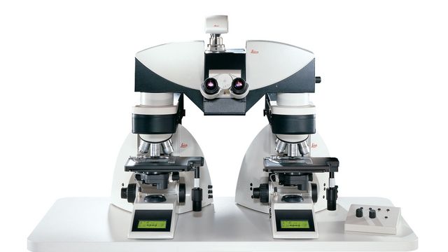
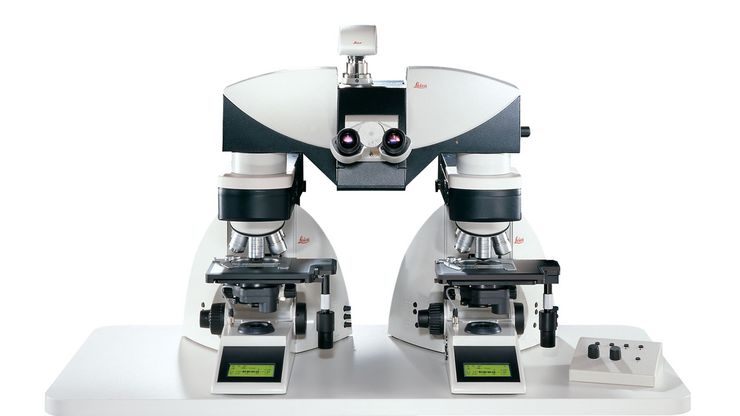
FS4000 LED
Forensic Comparison Microscope with LED Illumination
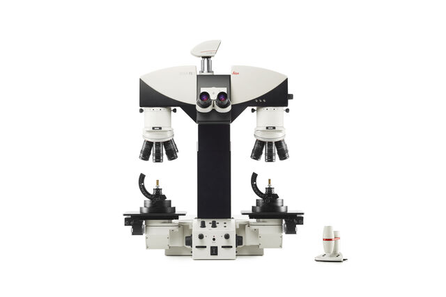
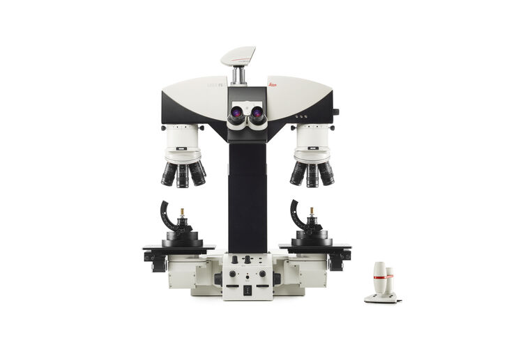
FS C & FS M
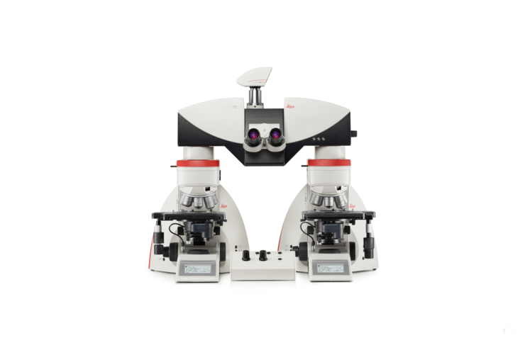
Comparison macroscopes for examination of forensic evidence
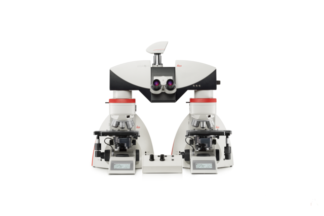
FS CB
Motorized Forensic Comparison Microscope
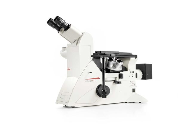
Leica DMi8 M / C / A
Inverted Microscopes for Industry
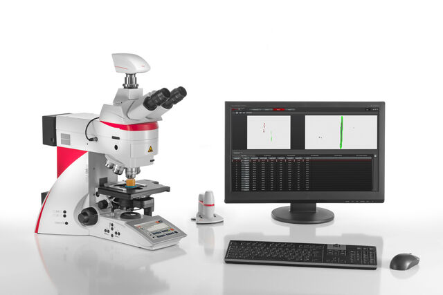
LAS X Steel Expert
Steel Analysis Software
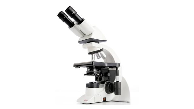
DM1000 LED
Uniquely ergonomic system microscope with LED illumination
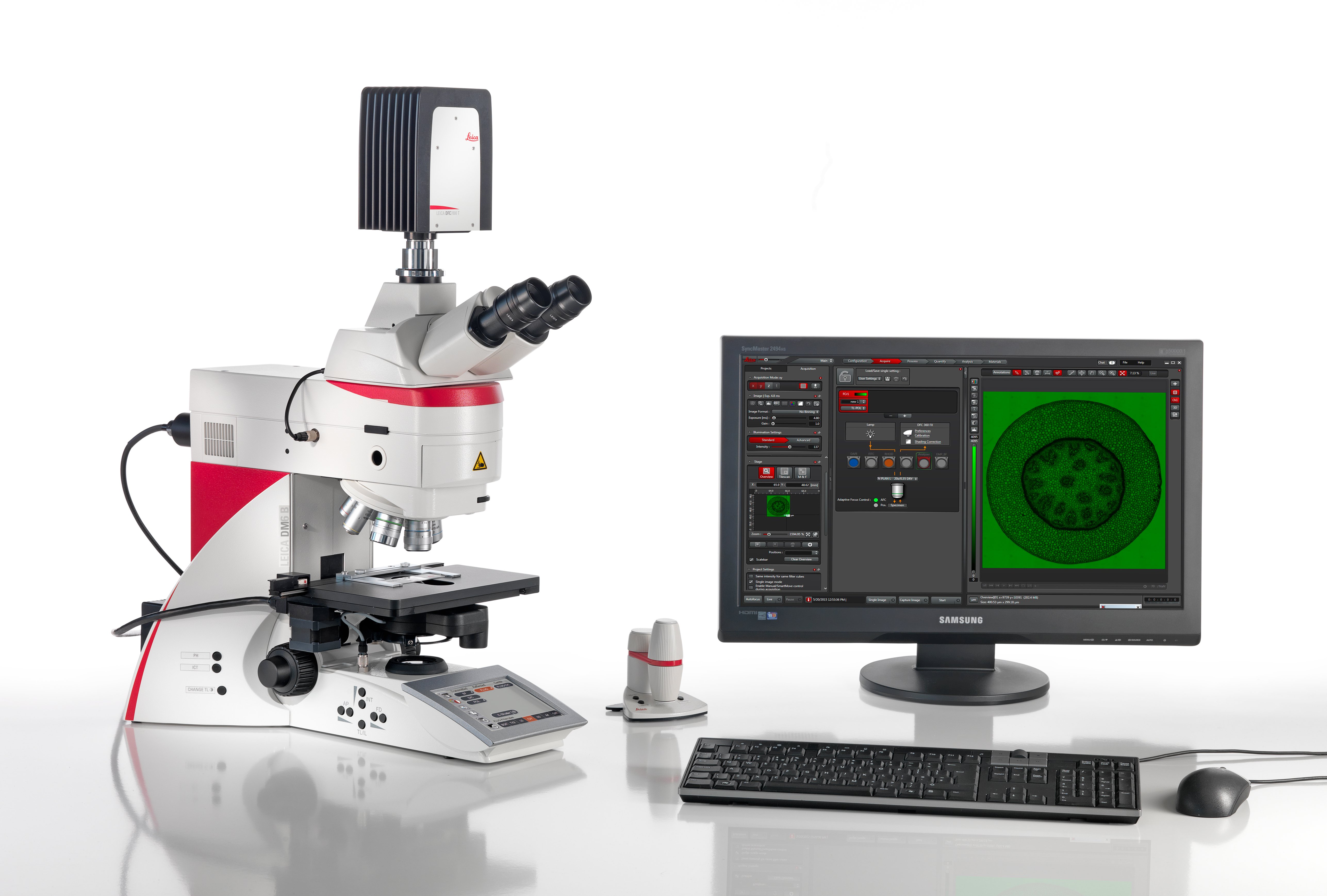
LAS X Widefield Systems
Fluorescence Microscope System for Advanced Imaging and Analysis
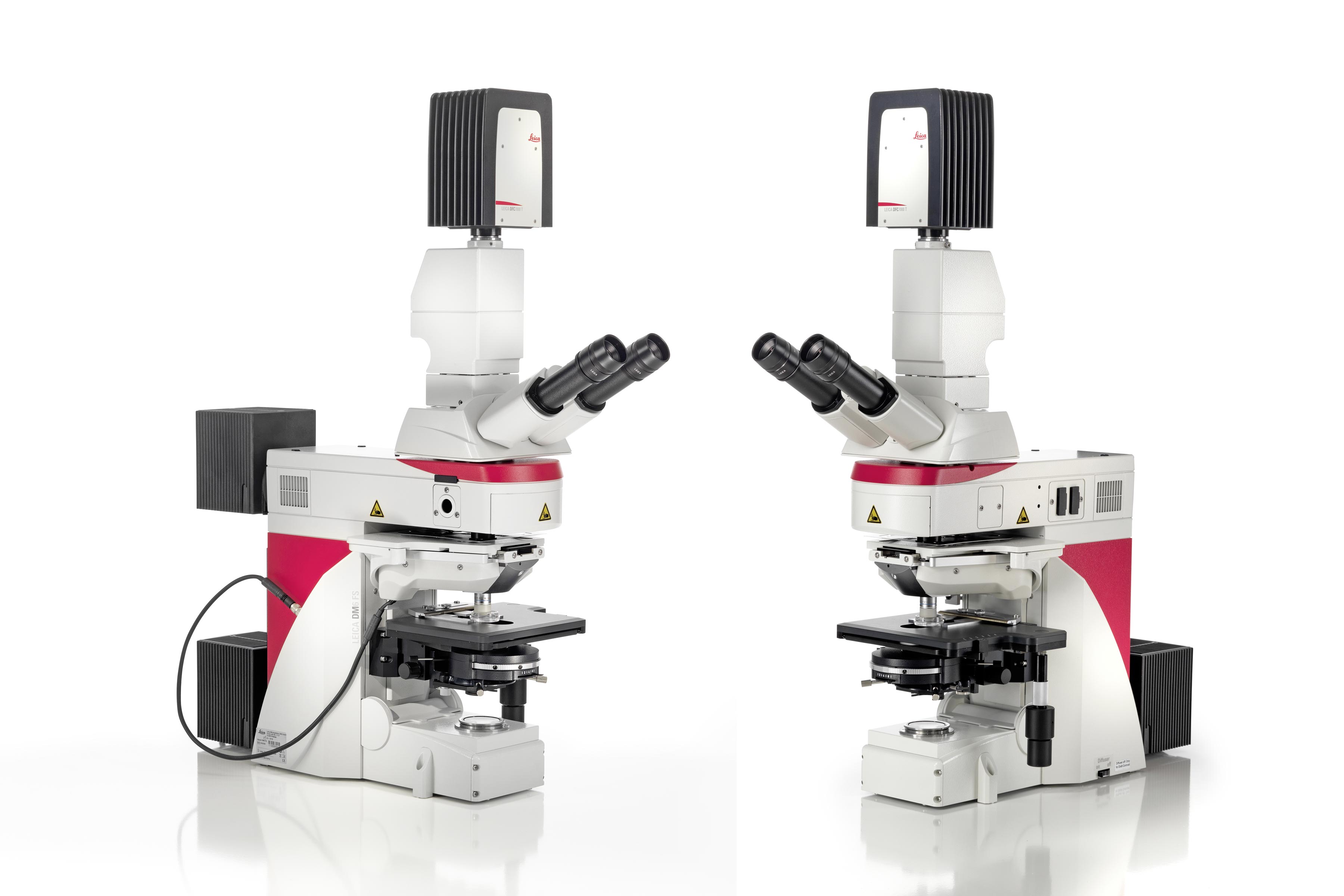
DM6 FS
Fixed Stage Microscope for Electrophysiology and in vivo Imaging
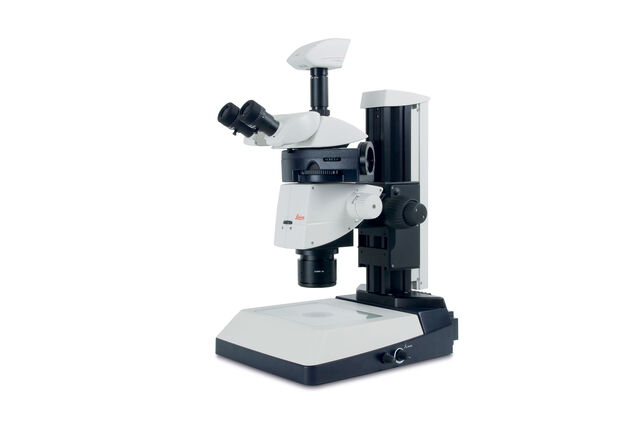
M165 FC
Fluorescent stereomicroscope
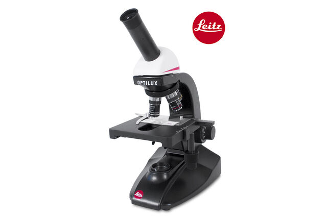
Leitz Optilux
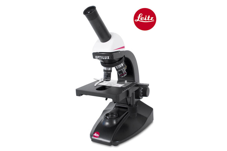
View the Finest Details - Compound Microscope
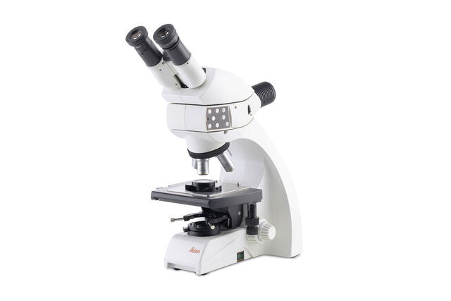
DM750 M
Binocular materials microscope for education, basic metallography, and forensics education
Leica Science Lab Show subnavigation
Read our latest articles about Light Microscopes
The knowledge portal of Leica Microsystems offers scientific research and teaching material on the subjects of microscopy. The content is designed to support beginners, experienced practitioners and scientists alike in their everyday work and experiments.
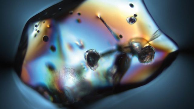
Ensuring Glass Quality with the Polarization Microscopy Advantage
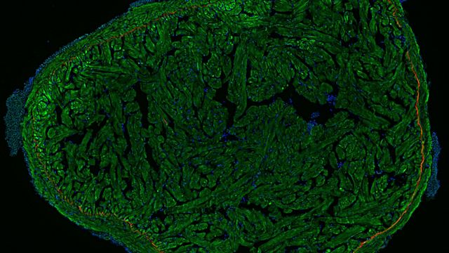
A Guide to Fluorescence Microscopy
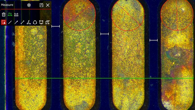
How to Select the Right Measurement Microscope
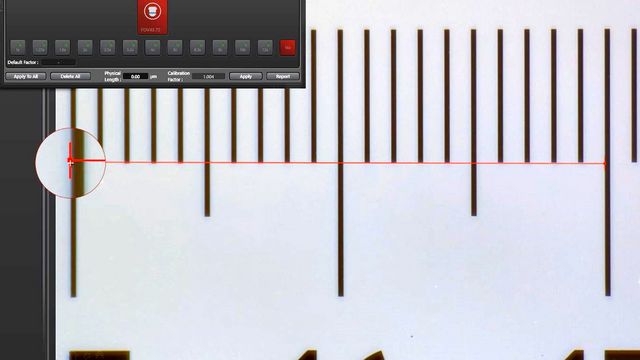
Microscope Calibration for Measurements: Why and How You Should Do It
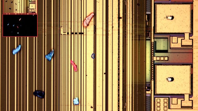
Visualizing Photoresist Residue and Organic Contamination on Wafers
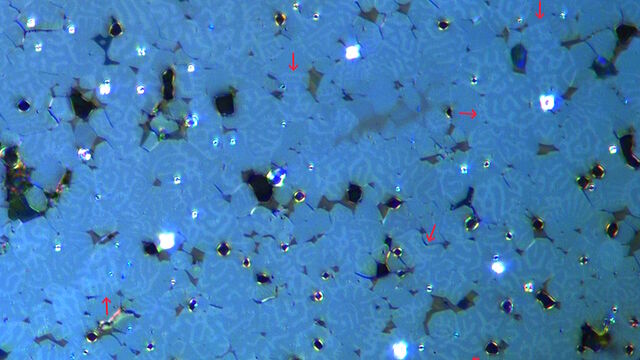
Rapidly Visualizing Magnetic Domains in Steel with Kerr Microscopy

6-Inch Wafer Inspection Microscope for Reliably Observing Small Height Differences
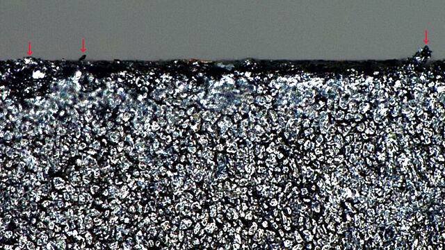
Burr Detection During Battery Manufacturing
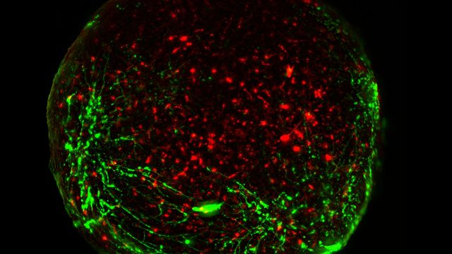
Guide to Live-Cell Imaging
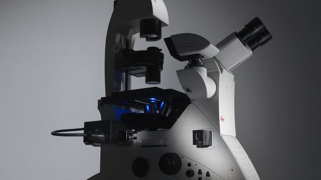
Factors to Consider When Selecting a Research Microscope
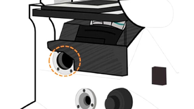
Infinity Optical Systems - From “Infinity Optics” to the Infinity Port

AI meets Deep Visual Proteomics (DVP) to Advance Disease Research
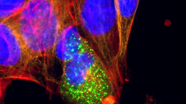
Microscopy and AI Solutions for 2D Cell Culture
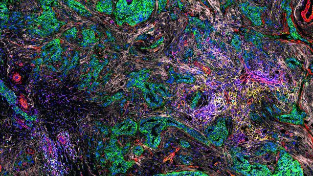
AI-Powered Hi-Plex Spatial Analysis Tools for Breast Cancer Research
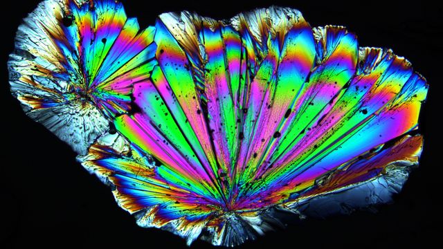
Polarizing Microscope Image Gallery
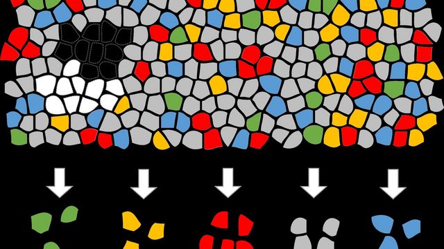
Biomarker Discovery with Laser Microdissection
Key Questions
1What is your application?
The Leica compound light microscope you need depends on your application. The demands of different applications are best met by different light microscopes and light microscope parts. The modular design of Leica light microscopes enable a customized solution to be set up for a specific application.
2Which type of samples do you need to visualize?
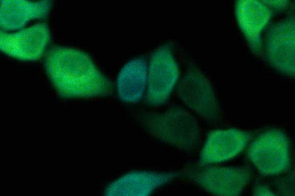
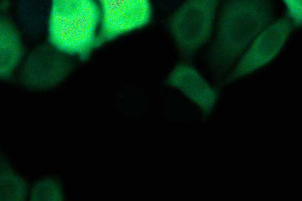
Whether you are observing samples for routine laboratory work, materials production or analysis, or complex life science research, the optical resolution, contrast, depth of field, and image quality you need are provided by Leica light microscope solutions. Additionally, light microscope parts and accessories, such as objectives, illumination types, and digital cameras, along with the Leica Application Suite software, can further customize and optimize the solution for your specific application needs.
3What about the budget for the light microscope solution?
Customized light microscope solutions can lead to higher investment costs, but they allow you and your colleagues to increase productivity. A microscope solution can be optimized essentially for almost any application due to a large variety of light microscopes, light microscope parts, and accessories.
4What is the difference between a compound light microscope and stereo microscope?
A compound light and stereo microscope both use optics with an objective lens and eyepieces or oculars for viewing the image of the sample. In general, a compound light microscope achieves a higher range of magnification than a stereo. There is only one light channel for viewing the sample with a compound microscope, so a 2D image is seen. A stereo microscope has two light channels, one for each eye of the observer, which enable a 3D image of the sample to be seen. If either a compound or stereo microscope is equipped with a camera, then just one light channel is used and only a 2D image can be recorded.

Meet Mica
The world’s first Microhub
Mica enables microscopy access for all, removes the constraints of traditional four-color fluorescence imaging and radically simplifies workflows.
Light Microscopes 45
DM4 B & DM6 B
M50, M60 & M80
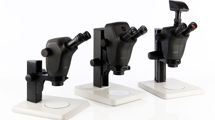
Ivesta 3


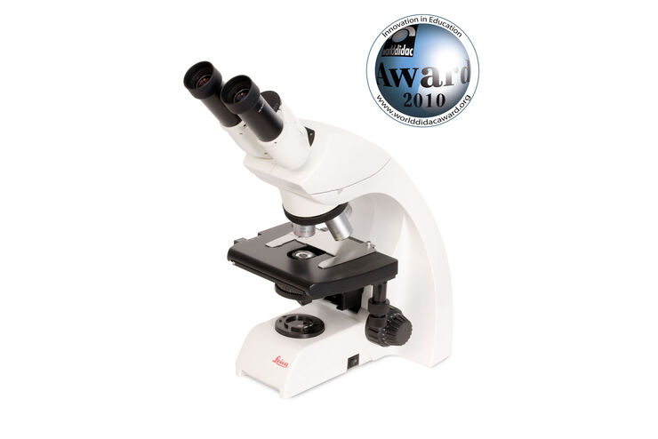
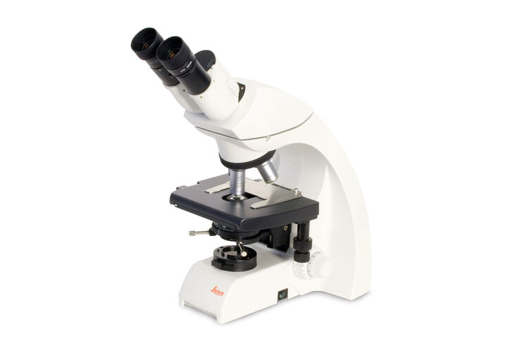
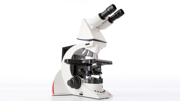
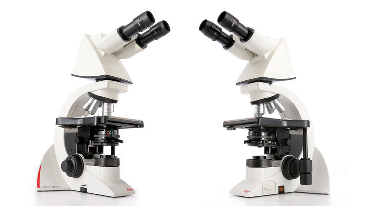
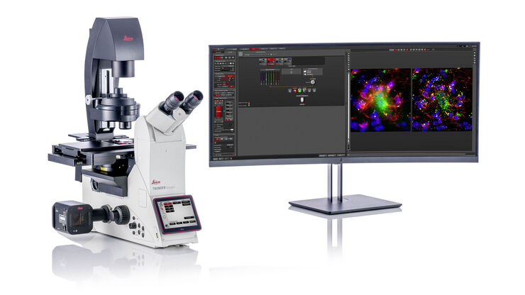
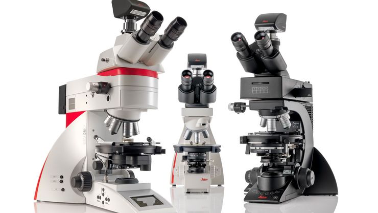
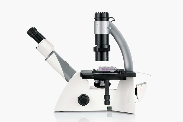
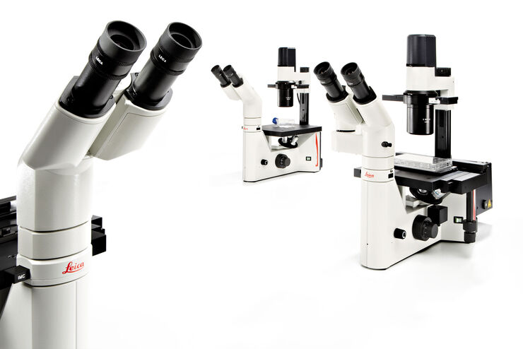
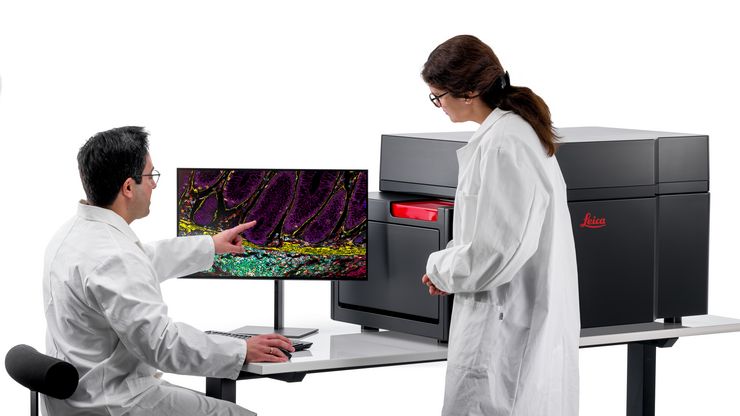
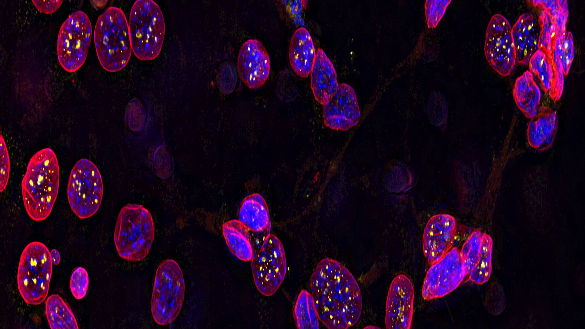
Cell Biology Research
Imaging solutions offered by Leica Microsystems are designed to maximize your cell biology research.
Follow us on Instagram
Frequently Asked Questions Light Microscopes
Leica microscopes are modular and shipped in the configuration that best fits your stated needs or application. In case your needs change later, you can always upgrade your workstation by adding available accessories.
Leica Microsystems offers the free Store & Recall software (available with the LAS X software platform for industry) which allows you to customize the microscope functions, adapting them to the requirements and needs of each individual user. The software also allows you to restore all system settings saved with the acquired image.
All our encoded microscope solutions offer calibration and image comparison. Furthermore, Leica Microsystems offers the free Store & Recall software which allows you to restore all system settings saved with the acquired image.
No, you don’t. By installing a FLEXACAM C1 camera, you can directly save the images on an IT network server or USB medium. You can also send the images via e-mail over your network without the need for a PC.
With the FLEXACAM C1 camera you can directly save images on an IT network server or USB medium. You can also send the images via e-mail without having to use a computer.
There are a lot of accessories. Please get in contact with your local Leica sales representative.
There are a lot of ergonomic accessories for Leica light microscopes. Please contact your local Leica sales representative for more details or visit: Ergonomic Accessories for Stereo Microscopes
LAS X software only runs with Windows, but for a MAC we have a dedicated software called Leica Acquire that you could download for free from the Apple store: https://apps.apple.com/it/app/leica-acquire/id733706983?mt=12
However, there is no software for Linux.
Yes, use of a 3rd party software is possible: https://www.splashtop.com/classroom
We have adapters for all C-mount compatible cameras.
The free AirLab software, compatible with iOS or ANDROID, allows the user to immediately share images, videos, and comments.
A compound microscope uses optics to produce a magnified image of a sample so that with details of it can be observed that are undetectable with the naked eye. The most basic optics of a compound microscope has at least 2 lenses: i) an objective placed nearby the sample which creates a magnified, real image of it and ii) eyepieces or oculars which are used to view the real image of the sample. A human who looks through the eyepieces sees the sample as a virtual image on his/her retina. For more information, please refer to the Science Lab article: Optical Microscopes – Some Basics
The maximum useful magnification value achieved with any type of light microscope depends on its ultimate resolving power or maximal resolution. The resolution depends on the microscope objective lens numerical aperture (NA). At low magnification values, the NA is small leading to a low resolution. At high magnification values, the NA is high yielding a high resolution. However, because the NA has a finite maximum value, approximately 1.3, the “useful” range of magnification is limited to about 1,800x for conventional light microscopes. Magnification beyond the useful range, which can occur whenever digital microscope cameras display images on large monitors, is called "empty” magnification. In this non-useful magnification range, the sample structures appear larger, but no additional details are resolved. For more information, please refer to the Science Lab articles: Beware of "Empty" Magnification, What Does 30,000:1 Magnification Really Mean?