
Science Lab
Science Lab
Willkommen auf dem Wissensportal von Leica Microsystems. Hier finden Sie wissenschaftliches Forschungs- und Lehrmaterial rund um das Thema Mikroskopie. Das Portal unterstützt Anfänger, erfahrene Praktiker und Wissenschaftler gleichermaßen bei ihrer täglichen Arbeit und ihren Experimenten. Erkunden Sie interaktive Tutorials und Anwendungshinweise, entdecken Sie die Grundlagen der Mikroskopie ebenso wie High-End-Technologien. Werden Sie Teil der Science Lab Community und teilen Sie Ihr Fachwissen.
Filter articles
Tags
Beitragstyp
Produkte
Loading...
 embedded in bulk vitreous ice (blue).")
Cryo-ET Sample Preparation: From Waffle Method to Serial Lift-Out
Cryo-ET sample preparation becomes more demanding when specimens are thicker, larger, or more complex. This webinar brings together four perspectives on how high-pressure freezing can be connected…
Loading...
 and phalloidin (magenta), imaged using Viventis SCAPE; scale bar 50μm. Courtesy of Marina Cuenca and Heleen Jungen (Dayton lab), EMBL Barcelona.")
What’s the Best Organoid Imaging Approach for Early Drug Discovery?
Organoids and other complex in vitro models (CIVMs) are becoming increasingly important in early drug discovery and translational research, driven by the need for more predictive, human-relevant data…
Loading...

Waffle Method Workflow: From HPF to Cryo-ET Lamellae
Waffle freezing provides an HPF-based route to cryo-ET sample preparation. This workflow guide follows the process from grid and carrier assembly to vitrification, cryo-FIB milling, lamella…
Loading...
 2D slices of a 1 mm diameter midbrain neural organoid stained with DAPI (blue, nuclear stain), β-tubulin (green, neuronal stain), and GFAP (red, astrocyte stain).")
Fast, High-Contrast Widefield Imaging of Optically Challenging Samples
Live‑cell imaging of large, complex biological samples often requires large fields of view, sub-cellular resolution, high-sensitivity, and fast acquisition – all while maintaining low illumination…
Loading...

Dental Loupes vs Microscopes: Exploring Visualization Options in Dentistry
Dental professionals often ask: “Should I use dental loupes or invest in a microscope?” This article explores the key differences between dental microscopes and dental loupes, focusing on…
Loading...

Spatial Proteomics Workflow in Blood Cancer (MPNs)
Megakaryocytes play a central role in the biology of myeloproliferative neoplasms (MPNs), yet their in vivo proteomic characterization remains a major challenge due to low abundance and disrupted…
Loading...
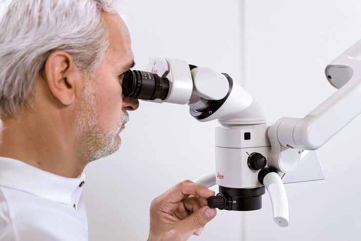
Sechs wichtige Faktoren für die Auswahl eines Dentalmikroskops
In der Zahnmedizin ist das Operationsmikroskop mittlerweile ein wichtiges Werkzeug für qualitativ hochwertige und erfolgreiche Operationen, insbesondere im Bereich der Endodontie. Ein Mikroskop…
Loading...

Multiscale Imaging of Organoids: High Content to Light Sheet
Learn multiscale organoid imaging: fixed high content phenotyping, gentle dual view light sheet, and reproducible pipelines that turn 3D data into insights.
Loading...
 and acceptor (A) molecule which participate in FRET (Förster resonance energy transfer).")
Was ist FRET mit FLIM (FLIM-FRET)?
Der Beitrag erläutert die FLIM-FRET-Methode, die Resonanzenergietransfer und Fluoreszenz-Lebensdauer-Imaging zur Untersuchung von Protein-Protein Wechselwirkungen kombiniert.
