
Filter articles
タグ
製品
Loading...
Workflows & Protocols: How to Use a Leica Laser Microdissection System and Qiagen Kits for Successful RNA Analysis
Laser Microdissection (LMD) allows isolating individual cells or chromosomes and is a well established technique for sample preparation prior downstream analysis of the nucleic acid content via PCR or…
Loading...

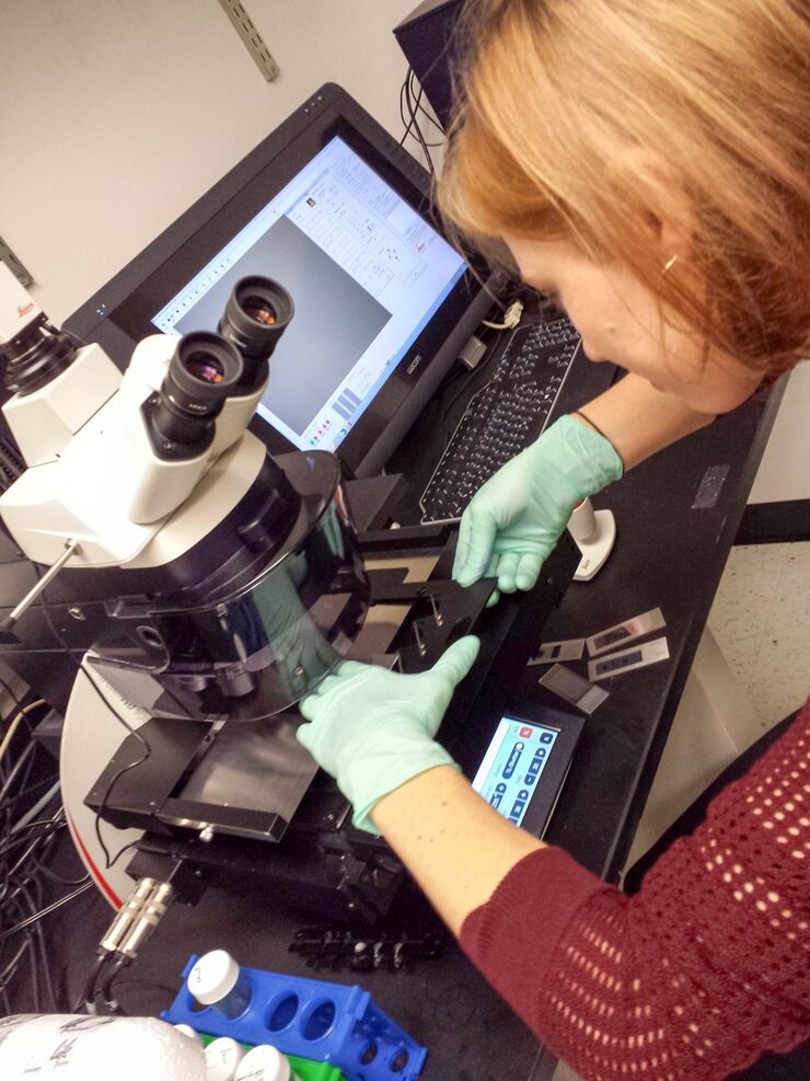
What You Always Wanted to Know About Digital Microscopy, but Never Got Around to Asking
Digital microscopy is one of the buzz words in microscopy – and there are a couple of facts that are useful to know. Georg Schlaffer, Product Manager with Leica Microsystems, has often been asked…
Loading...

Confocal and Light Sheet Imaging
Optical imaging instrumentation can magnify tiny objects, zoom in on distant stars and reveal details that are invisible to the naked eye. But it notoriously suffers from an annoying problem: the…
Loading...
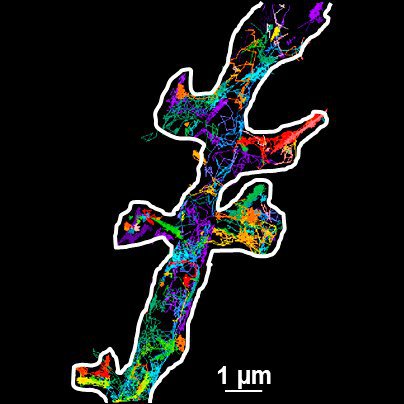
Universal PAINT – Dynamic Super-Resolution Microscopy
Super-resolution microscopy techniques have revolutionized biology for the last ten years. With their help cellular components can now be visualized at the size of a protein. Nevertheless, imaging…
Loading...

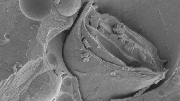
Immersion Freezing for Cryo-Transmission Electron Microscopy: Applications
A well established usage case for cryo-TEM is three-dimensional reconstruction of isolated macromolecules, virus particles, or filaments. These approaches are based on averaging of repetitive…
Loading...
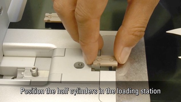
Video Tutorials: Filling and Assembling of Different Carriers for High-Pressure Freezing
High pressure freezing (HPF) is a cryo-fixation method primarily for biological samples, but also for a variety of non-biological materials. It is a technique that yields optimal preservation in many…
Loading...
Brief Introduction to Freeze Fracture and Etching
Freeze fracture describes the technique of breaking a frozen specimen to reveal internal structures. Freeze etching is the sublimation of surface ice under vacuum to reveal details of the fractured…
Loading...
Brief Introduction to Specimen Trimming
Before ultrathin sectioning a sample with an ultramicrotome it has to be pre-prepared. For this pre-preparation, special attention must be paid to the sample size (size of the section), location of…
Loading...
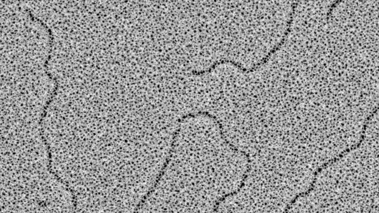
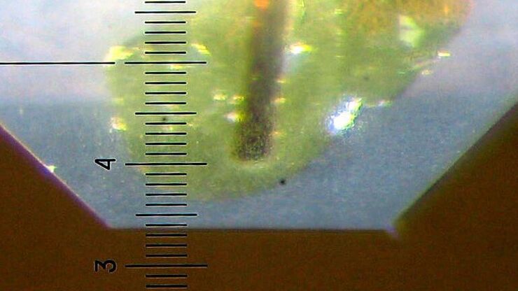
Glycerol Spraying/Platinum Low Angle Rotary Shadowing of DNA with the Leica EM ACE600 e-beam
Glycerol spraying/low angle rotary shadowing (Aebi and Baschong, 2006) is a preparation technique used in biology to visualize structures yielding insufficent contrast with other techniques, due to…
