
Science Lab
Science Lab
Learn. Share. Contribute. The knowledge portal of Leica Microsystems. Find scientific research and teaching material on the subject of microscopy. The portal supports beginners, experienced practitioners and scientists alike in their everyday work and experiments. Explore interactive tutorials and application notes, discover the basics of microscopy as well as high-end technologies. Become part of the Science Lab community and share your expertise.
Filter articles
Tags
Story Type
Products
Loading...

The “Waffle Method”: High-Pressure Freeze Complex Samples
This article describes the advantages of a special high pressure freezing method, the so-called “Waffle Method”. Learn how the “Waffle Method” uses EM grids as spacers for high-pressure freezing,…
Loading...

Mastering Polymer Sectioning with Helmut Gnaegi
When it comes to ultramicrotomy, few names carry the weight of Helmut Gnaegi. As co-founder of Diatome, a global leader in diamond knife technology, Helmut has spent decades refining the art and…
Loading...
.")
How Fluorescence Guides Sectioning of Resin-embedded EM Samples
Electron microscopes, including transmission electron microscopes (TEM) and scanning electron microscopes (SEM), are widely utilized to gain detailed structural information about biological samples or…
Loading...

How to Save Time and Samples by Automated Ultramicrotomy
This article describes how 3D micro-CT data of a resin-embedded electron microscopy sample can be used to trim the specimen down to a defined target plane prior to sectioning. The interactive and…
Loading...
 and oblique (right) brightfield illumination using a Leica compound microscope. The defect on the wafer surface is clearly more visible with oblique illumination.")
Rapid Semiconductor Inspection with Microscope Contrast Methods
Semiconductor inspection during the production of patterned wafers and ICs (integrated circuits) is important for identifying and minimizing defects. To increase the efficiency of quality control in…
Loading...
. The grains are highlighted with arbitrary colors.")
High-Quality EBSD Sample Preparation
This article describes a method for EBSD sample preparation of challenging materials. The high-quality samples required for electron backscatter diffraction are prepared with broad ion-beam milling.
Loading...
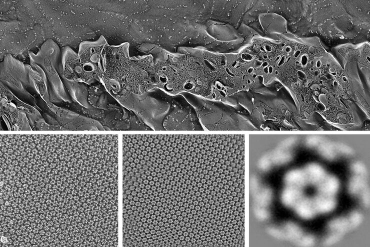
Advancing Cellular Ultrastructure Research
Freeze-fracture and freeze-etching are useful tools for studying flexible membrane-associated structures such as tight junctions or the enteric glycocalyx. Freeze-fracture and etching are two…
Loading...
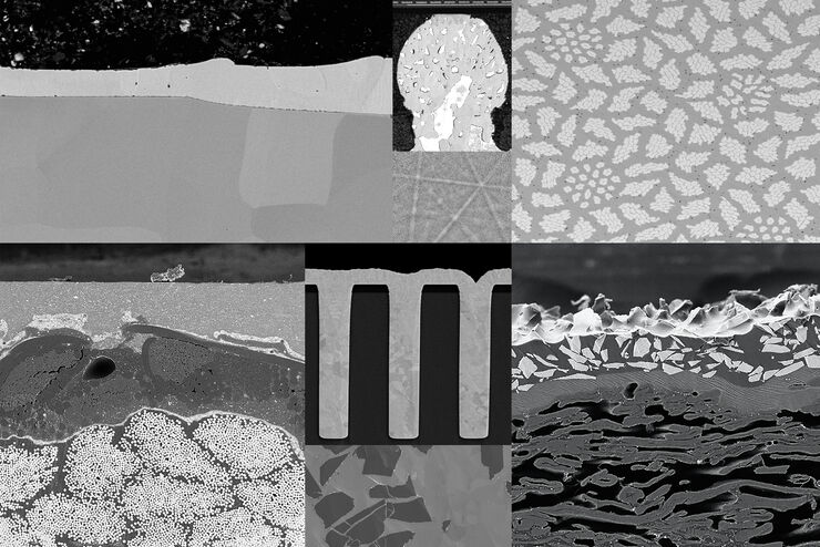
Ion Beam Milling Guide: Enhancing Surface Quality for High-Resolution Imaging and Analysis
In this article you can learn how to optimize the preparation quality of your samples by using the ion beam etching method with the EM TIC 3X ion beam milling machine. A short introduction of the…
Loading...
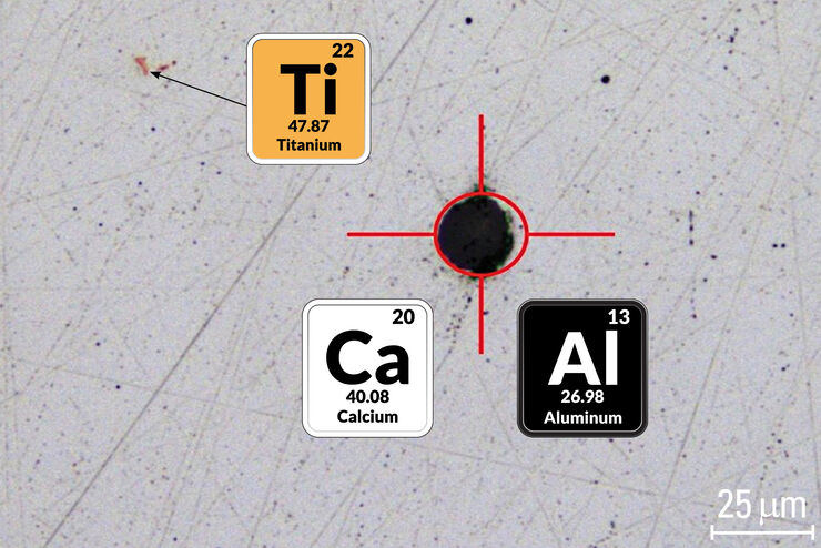
Visual and Chemical Analysis of Steel Microstructure: Faster Rating of Steel Quality
Simultaneous visual and chemical analysis of steel non-metallic inclusions with a 2-methods-in-1 solution, using optical microscopy and laser induced breakdown spectroscopy (LIBS), is described in…
