
Science Lab
Science Lab
Learn. Share. Contribute. The knowledge portal of Leica Microsystems. Find scientific research and teaching material on the subject of microscopy. The portal supports beginners, experienced practitioners and scientists alike in their everyday work and experiments. Explore interactive tutorials and application notes, discover the basics of microscopy as well as high-end technologies. Become part of the Science Lab community and share your expertise.
Filter articles
Tags
Story Type
Products
Loading...

Extended Live-cell Imaging at Nanoscale Resolution
Extended live-cell imaging with TauSTED Xtend. Combined spatial and lifetime information allow super-resolution microscopy at extremely low light dose.
Loading...
, actin network (ATTO 647N), and nuclear pore basket (CF 680R).")
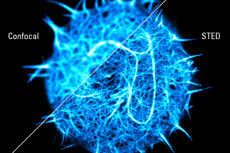
The Guide to STED Sample Preparation
This guide is intended to help users optimize sample preparation for stimulated emission depletion (STED) nanoscopy, specifically when using the STED microscope from Leica Microsystems. It gives an…
Loading...

Five-color FLIM-STED with One Depletion Laser
Webinar on five-color STED with a single depletion laser and fluorescence lifetime phasor separation.
Loading...

A Versatile Palette of Fluorescent Probes
Researchers at the Max Planck Institute for Medical Research in Heidelberg have developed a general strategy to synthesize live-cell compatible fluorogenic probes, and the result are the new MaP (Max…
Loading...

Benefits of Combining STED and Lifetime
In this interview, Professor Alberto Diaspro talks about the advantages of the White Light Laser and the TauSTED capabilities of STELLARIS 8 STED. He speaks about his experience with the confocal…
Loading...
, and trafficking vesicles labeled with CF594 (cyan - Biotium).")
A Guide to Super-Resolution
Find out more about Leica super-resolution microscopy solutions and how they can empower you to visualize in fine detail subcellular structures and dynamics.
Loading...
Super-resolved STED spectroscopy
Molecular interactions are key in cellular signalling. They are often ruled or rendered by the mobility of the involved molecules.
