
Science Lab
Science Lab
Learn. Share. Contribute. The knowledge portal of Leica Microsystems. Find scientific research and teaching material on the subject of microscopy. The portal supports beginners, experienced practitioners and scientists alike in their everyday work and experiments. Explore interactive tutorials and application notes, discover the basics of microscopy as well as high-end technologies. Become part of the Science Lab community and share your expertise.
Filter articles
Tags
Story Type
Products
Loading...

How FLIM Microscopy Helps to Detect Microplastic Pollution
The use of autofluorescence in biological samples is a widely used method to gain detailed knowledge about systems or organisms. This property is not only found in biological systems, but also…
Loading...

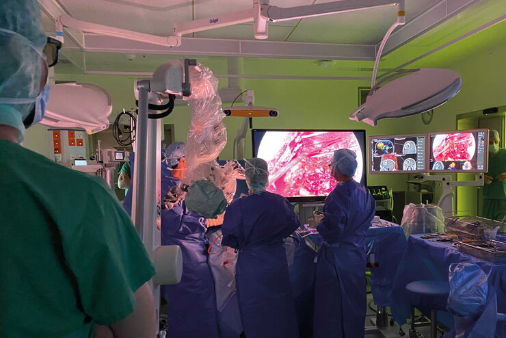
Oncological Reconstructive Surgery with the Leica M530 OHX Microscope
Precision is essential in oncological reconstructive surgery, in particular when it relies on free flap techniques. Microsurgical microscopes provide optimal visualization and help streamline the…
Loading...

Oncological Reconstructive Surgery: Why Use a Microscope
Recent advances in microsurgery are enhancing breast reconstruction for oncology patients, allowing both functional and aesthetic rehabilitation. More and more surgeons are adopting surgical…
Loading...
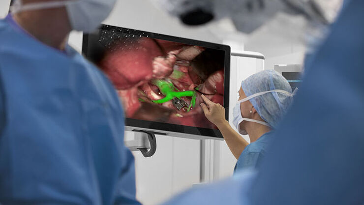
Neurosurgery with Heads-up Display
In the following video interviews Prof. Dr. Raphael Guzman, Vice Chairman of the Department of Neurosurgery at the University Hospital in Basel, Switzerland, talks about his experience in heads-up…
Loading...
Augmented Reality (AR) Fluorescence Image Gallery
Building on a decade of leadership in fluorescence imaging technology, GLOW800 AR fluorescence is the first of many modalities based on the proprietary GLOW AR platform.
The sophisticated imaging…
Loading...
Clinical Uses in Cerebrovascular and Skull Base Neurosurgery
In this webinar Dr. Bendok and Dr. Morcos explain how Augmented Reality and Fluorescence can enhance visualization and support surgical decision making. They present first-hand experience of the GLOW…
Loading...
 from the desired fluorescent signal from the cell membrane (green).")
Learn how to Remove Autofluorescence from your Confocal Images
Autofluorescence can significantly reduce what you can see in a confocal experiment. This article explores causes of autofluorescence as well as different ways to remove it, from simple media fixes to…
Loading...
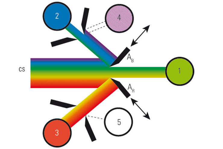
What is a Spectral Detector (SP Detector)?
The SP detector from Leica Microsystems denotes a compound detection unit for point scanning microscopes, in particular confocal microscopes. The SP detector splits light into up to 5 spectral bands.…
Loading...

Which Sensor is the Best for Confocal Imaging?
The Hybrid Photodetectors (HyD) are! Why that is the case is explained in this short Science Lab article.
