
Science Lab
Science Lab
Learn. Share. Contribute. The knowledge portal of Leica Microsystems. Find scientific research and teaching material on the subject of microscopy. The portal supports beginners, experienced practitioners and scientists alike in their everyday work and experiments. Explore interactive tutorials and application notes, discover the basics of microscopy as well as high-end technologies. Become part of the Science Lab community and share your expertise.
Filter articles
Tags
Story Type
Products
Loading...

The Power HyD Detector Family
Powerful photon counting detectors on the STELLARIS confocal platform provide improved photon counting, ultra-sensitive imaging and more color options in the NIR spectrum.
Loading...
 from the desired fluorescent signal from the cell membrane (green).")
Learn how to Remove Autofluorescence from your Confocal Images
Autofluorescence can significantly reduce what you can see in a confocal experiment. This article explores causes of autofluorescence as well as different ways to remove it, from simple media fixes to…
Loading...

Zebrafish Brain - Whole Organ Imaging at High Resolution
Structural information is key when one seeks to understand complex biological systems, and one of the most complex biological structures is the vertebrate central nervous system. To image a complete…
Loading...

What is a Resonant Scanner?
A resonant scanner is a type of galvanometric mirror scanner that allows fast image acquisition with single-point scanning microscopes (true confocal and multiphoton laser scanning). High acquisition…
Loading...
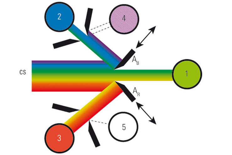
What is a Spectral Detector (SP Detector)?
The SP detector from Leica Microsystems denotes a compound detection unit for point scanning microscopes, in particular confocal microscopes. The SP detector splits light into up to 5 spectral bands.…
Loading...

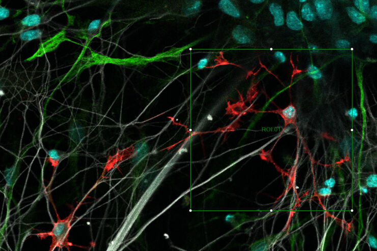
Improve 3D Cell Biology Workflow with Light Sheet Microscopy
Understanding the sub-cellular mechanisms in carcinogenesis is of crucial importance for cancer treatment. Popular cellular models comprise cancer cells grown as monolayers. But this approach…
Loading...
Resolved Field Number (RFN)
The field number (FN) for optical microscopes indicates the field of view (FOV). It corresponds to the area in the intermediate image that is observable through the eyepieces. Although, we cannot…
Loading...
What is a Field-of-View Scanner?
A field-of-view scanner is an assembly of galvanometric scanning mirrors used in single-point confocal microscopes that offer the correct optical recording of large field sizes. The field-of-view…
Loading...

What is a Tandem Scanner?
A Tandem Scanner is an assembly of two different types of scanning together in one system for true confocal point scanning. The Tandem Scanner consists of a three-mirror scanning base with the…
