
Science Lab
Science Lab
Learn. Share. Contribute. The knowledge portal of Leica Microsystems. Find scientific research and teaching material on the subject of microscopy. The portal supports beginners, experienced practitioners and scientists alike in their everyday work and experiments. Explore interactive tutorials and application notes, discover the basics of microscopy as well as high-end technologies. Become part of the Science Lab community and share your expertise.
Filter articles
Tags
Story Type
Products
Loading...

How to Streamline Your Histology Workflows
Streamline your histology workflows. The unique Fluosync detection method embedded into Mica enables high-res RGB color imaging in one shot.
Loading...

Accelerating Discovery for Multiplexed Imaging of Diverse Tissues
Explore IBEX: Open-source multiplexed imaging. Join the collaborative IBEX Imaging Community for optimized tissue processing, antibody selection, and human atlas construction.
Loading...

Notable AI-based Solutions for Phenotypic Drug Screening
Learn about notable optical microscope solutions for phenotypic drug screening using 3D-cell culture, both planning and execution, from this free, on-demand webinar.
Loading...

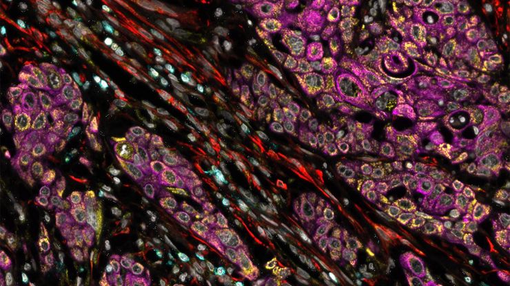
Understanding Tumor Heterogeneity with Protein Marker Imaging
Explore tumor heterogeneity and immune cell dynamics. See how quantitative imaging analysis reveals spatial relationships and molecular insights crucial for advancing cancer research and therapeutics.
Loading...

In Situ Identification of Cancer Stem Cell Niches in Hepatocellular Carcinoma
Discover how multiplexed imaging technology uncovers cancer stem cell niches in Hepatocellular Carcinoma using multiplex immunodetection, revealing extracellular matrix dynamics. Explore precise…
Loading...

Multiplexing with Luke Gammon: Advance your Spatial Biology Research
Learn how multiplexing imaging and spatial biology can help researchers better understand complex biological systems. In this interview, Dr. Gammon and Dr. Pointu of Leica Microsystems discuss pain…
Loading...

Differential Interference Contrast (DIC) Microscopy
This article demonstrates how differential interference contrast (DIC) can be actually better than brightfield illumination when using microscopy to image unstained biological specimens.
Loading...
Cell DIVE: Unraveling Pathways in Pancreatic Cancer
Unlock new insights into tumour-immune interactions with spatial biology. Join our webinar to learn how to label >60 biomarkers per tissue section and use advanced clustering and analysis techniques…
Loading...

How to Determine Cell Confluency with a Digital Microscope
This article shows how to measure cell confluency in an easy and consistent way with Mateo TL, increasing confidence in downstream experiments.
