
Science Lab
Science Lab
Learn. Share. Contribute. The knowledge portal of Leica Microsystems. Find scientific research and teaching material on the subject of microscopy. The portal supports beginners, experienced practitioners and scientists alike in their everyday work and experiments. Explore interactive tutorials and application notes, discover the basics of microscopy as well as high-end technologies. Become part of the Science Lab community and share your expertise.
Filter articles
Tags
Story Type
Products
Loading...

TauInteraction – Studying Molecular Interactions with TauSense
Fluorescence microscopy constitutes one of the pillars in life sciences and is a tool commonly used to unveil cellular structure and function. A key advantage of fluorescence microscopy resides in the…
Loading...
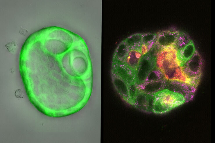
How Does The Cytoskeleton Transport Molecules?
VIDEO ON DEMAND - See how 3D cysts derived from MDCK cells help scientists understand how proteins are transported and recycled in tissues and the role of the cytoskeleton in this transport.
Loading...
 embryo, from sphere stage to somite stages.")
Studying Early Phase Development of Zebrafish Embryos
This video on demand focuses on combining widefield and confocal imaging to study the early-stage development of zebrafish embryos (Danio rerio), from oocyte to multicellular stage.
Loading...

How To Get Multi Label Experiment Data With Full Spatiotemporal Correlation
This video on demand focuses on the special challenges of live cell experiments. Our hosts Lynne Turnbull and Oliver Schlicker use the example of studying the mitochondrial activity of live cells.…
Loading...

Find Relevant Specimen Details from Overviews
Switch from searching image by image to seeing the full overview of samples quickly and identifying the important specimen details instantly with confocal microscopy. Use that knowledge to set up…
Loading...

How to Target Fluorescent Structures in 3D for Cryo-FIB Milling
This article describes the major steps of the cryo-electron tomography workflow including super-resolution cryo-confocal microscopy. We describe how subcellular structures can be precisely located in…
Loading...

Precise 3D Targeting for EM Imaging - Access What Matters
Find out how the seamless cryo-electron tomography workflow Coral Cryo uses confocal super resolution to target your structure of interest more precisely.
Loading...
, unsaturated lipids (magenta, 3050 cm-1), collagen (SHG, cyan). Sample courtesy of R. Rudolf, J Klicks, Hochschule Mannheim")
The Potential of Coherent Raman Scattering Microscopy at a Glance
Coherent Raman scattering microscopy (CRS) is a powerful approach for label-free, chemically specific imaging. It is based on the characteristic intrinsic vibrational contrast of molecules in the…
Loading...

Simplifying Complex Fluorescence Multiwell Plate Assays
Apoptosis, or programmed cell death, occurs during organism embryo development to eliminate unwanted cells and during healing in adults to rid the body of damaged cells and help prevent cancer.…
