
Science Lab
Science Lab
Learn. Share. Contribute. The knowledge portal of Leica Microsystems. Find scientific research and teaching material on the subject of microscopy. The portal supports beginners, experienced practitioners and scientists alike in their everyday work and experiments. Explore interactive tutorials and application notes, discover the basics of microscopy as well as high-end technologies. Become part of the Science Lab community and share your expertise.
Filter articles
Tags
Story Type
Products
Loading...

A Guide to Model Organisms in Research
A model organism is a species used by researchers to study specific biological processes. They have similar genetic characteristics to humans and are commonly used in research areas such as genetics,…
Loading...

Guide to Microscopy in Cancer Research
Cancer is a complex and heterogeneous disease caused by cells deficient in growth regulation. Genetic and epigenetic changes in one or a group of cells disrupt normal function and result in…
Loading...

Introduction to Widefield Microscopy
This article gives an introduction to widefield microscopy, one of the most basic and commonly used microscopy techniques. It also shows the basic differences between widefield and confocal…
Loading...
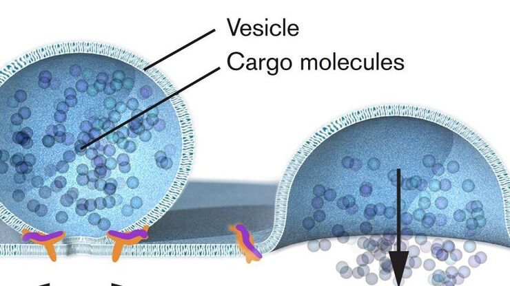
Nobel Prize 2013 in Physiology or Medicine for Discoveries of the Machinery Regulating Vesicle Traffic
On October 7th 2013, The Nobel Assembly at Karolinska Institutet has decided to award The Nobel Prize in Physiology or Medicine 2012 jointly to James E. Rothman, Randy W. Schekman and Thomas C. Südhof…
Loading...

Nobel Prize 2012 in Physiology or Medicine for Stem Cell Research
The Nobel Prize recognizes two scientists who discovered that mature, specialised cells can be reprogrammed to become immature cells capable of developing into all tissues of the body. Their findings…
Loading...
 inoculated with cowpea mosaic virus (CPMV) containing the GFP-gene inserted between the movement protein (MP) and the capsid proteins (CPs) in the viral RNA 2")
Introduction to Live-Cell Imaging
The understanding of complex and fast cellular dynamics is an important step to get insight into biological processes. Therefore, today’s life science research more and more demands studying…
