
Science Lab
Science Lab
Learn. Share. Contribute. The knowledge portal of Leica Microsystems. Find scientific research and teaching material on the subject of microscopy. The portal supports beginners, experienced practitioners and scientists alike in their everyday work and experiments. Explore interactive tutorials and application notes, discover the basics of microscopy as well as high-end technologies. Become part of the Science Lab community and share your expertise.
Filter articles
Tags
Story Type
Products
Loading...

How did Laser Microdissection enable Pioneering Neuroscience Research?
Dr. Marta Paterlini, a Senior Scientist at the Karolinska Institute, shares her experience of using laser microdissection (LMD) in groundbreaking research into adult human neurogenesis and offers…
Loading...

Buying an Ophthalmic Microscope? Gain Peers Insights from Dr. Dhami
In this article, learn how Dr. Abhinav Dhami, an ophthalmic surgery consultant in North India, enhances his surgical precision using the M822 ophthalmic microscope from Leica Microsystems and the key…
Loading...
 set-up.")
Augmented Reality: Transforming Neurosurgical Procedures
In this ebook, you will explore the exciting advances that Augmented Reality (AR) brings to the field of neurosurgery. This comprehensive guide, including explanatory videos, addresses key questions…
Loading...
 tissue.")
AI-Powered Multiplexed Image Analysis to Explore Colon Adenocarcinoma
In this application note, we demonstrate a spatial biology workflow via an AI-powered multiplexed image analysis-based exploration of the tumor immune microenvironment in colon adenocarcinoma.
Loading...

Laser Microdissection Protocols for Tissue and Cell Isolation - Download free eBook
In this Bio-protocol Selections, we present a collection of open-access, detailed methods papers using LCM to purify and isolate tissues and cells from plants, mouse embryos, cancer cells, neurons,…
Loading...

How do Cells Talk to Each Other During Neurodevelopment?
Professor Silvia Capello presents her group’s research on cellular crosstalk in neurodevelopmental disorders, using models such as cerebral organoids and assembloids.
Loading...

Tympanoplasty Surgery: Optimal Approaches and Tools
Discover tympanoplasty surgery case studies illustrating the standard approaches: post-auricular, endaural and transcanal. Gain insights from Dr. Flanagan on selecting the adequate surgical approach…
Loading...
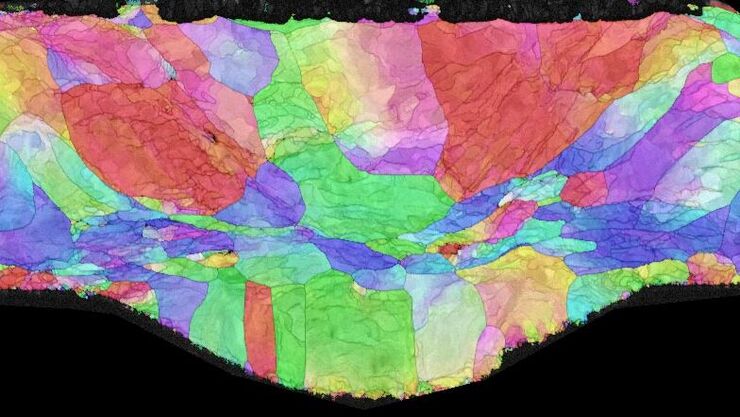
Workflow Solutions for Sample Preparation Methods for Material Science
This brochure presents and explains appropriate workflow solutions for the most frequently required sample preparation methods for material science samples.
Loading...
.")
Dual-View LightSheet Microscope for Large Multicellular Systems
Visualizing the dynamics of complex multicellular systems is a fundamental goal in biology. To address the challenges of live imaging over large spatiotemporal scales, Franziska Moos et. al. present…
