

Science Lab
Science Lab
Learn. Share. Contribute. The knowledge portal of Leica Microsystems. Find scientific research and teaching material on the subject of microscopy. The portal supports beginners, experienced practitioners and scientists alike in their everyday work and experiments. Explore interactive tutorials and application notes, discover the basics of microscopy as well as high-end technologies. Become part of the Science Lab community and share your expertise.
Filter articles
Tags
Story Type
Products
Loading...

The Principles of White Light Laser Confocal Microscopy
The perfect light source for confocal microscopes in biomedical applications has sufficient intensity, tunable color and is pulsed for use in lifetime fluorescence. Furthermore, it should offer means…
Loading...

Introduction to Live-Cell Imaging
The understanding of complex and fast cellular dynamics is an important step to get insight into biological processes. Therefore, today’s life science research more and more demands studying…
Loading...
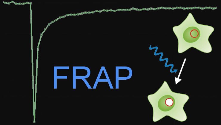
Fluorescence Recovery after Photobleaching (FRAP) and its Offspring
FRAP (Fluorescence recovery after photobleaching) can be used to study cellular protein dynamics: For visualization the protein of interest is fused to a fluorescent protein or a fluorescent dye. A…
Loading...
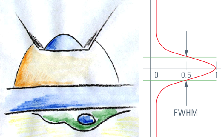
Confocal Optical Section Thickness
Confocal microscopes are employed to optically slice comparably thick samples.


