

Science Lab
Science Lab
Learn. Share. Contribute. The knowledge portal of Leica Microsystems. Find scientific research and teaching material on the subject of microscopy. The portal supports beginners, experienced practitioners and scientists alike in their everyday work and experiments. Explore interactive tutorials and application notes, discover the basics of microscopy as well as high-end technologies. Become part of the Science Lab community and share your expertise.
Filter articles
Tags
Story Type
Products
Loading...
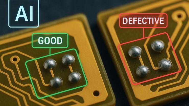
AI Image Analysis for Reliable Defect Detection
AI-based visual inspection with microscopy helps make quality control in industrial environments efficient by reducing manual inspection effort concerning defect detection, classification, and…
Loading...

An Overview of Artificial Intelligence (AI) and Machine Learning
AI is rapidly changing industrial inspection, quality control, and manufacturing workflows. But not all AI technologies work the same way. From machine learning and deep learning to the newer concept…
Loading...

Why the Future of Microscopic Inspection Is AI-Assisted, Not Fully Automated
The next wave of QC productivity gains will not come from building more automated inspection lines. Instead, it will come from augmenting millions of inspection decisions that are made manually every…
Loading...
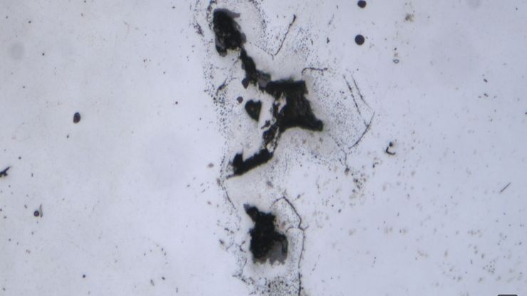
AI-Based Particle Counting with the Aivaro AI Feature Detection Software Module
The Aivaro AI Feature Detection software module enables fast manual microscope inspection of particulate contamination on various material samples. By training different classes, reflective particles,…
Loading...
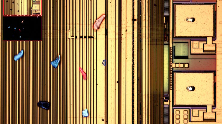
Visualizing Photoresist Residue and Organic Contamination on Wafers
As the scale of integrated circuits (ICs) on semiconductors passes below 10 nm, efficient detection of organic contamination, like photoresist residue, and defects during wafer inspection is becoming…
Loading...
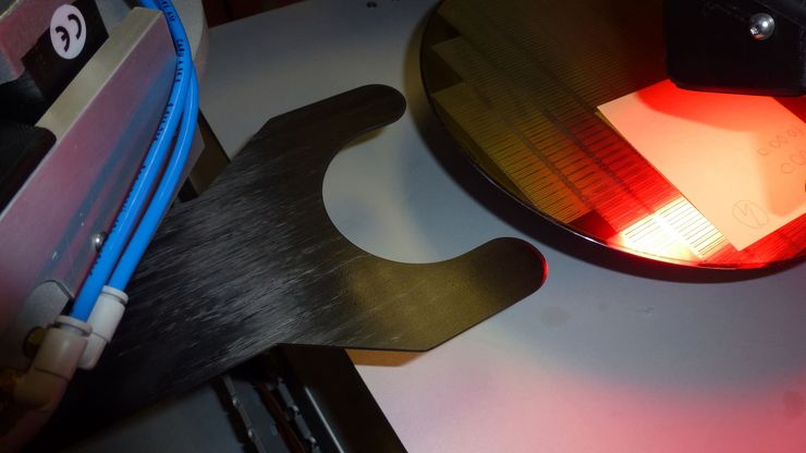
Safe Wafer Loading for Microscope Inspection without Hand Contact
How automated silicon wafer loading for microscope inspection helps improve microelectronics process control and production efficiency is explained in this article. Manual handling of wafers has a…
Loading...

Burr Detection During Battery Manufacturing
See how optical microscopy can be used for burr detection on battery electrodes and determination of damage potential to achieve rapid and reliable quality control during battery manufacturing.
Loading...

Quality Assurance Improvement Across Industries
Precision is paramount. Imagine a pacemaker that fails mid-operation or a semiconductor flaw that causes a critical system crash. In industries, such as medical devices, electronics, and…
Loading...
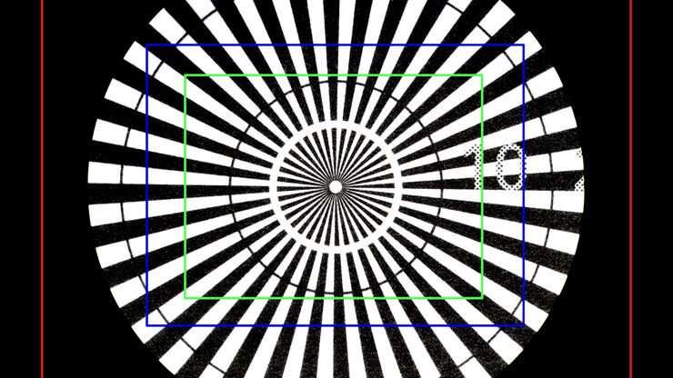
Understanding Clearly the Magnification of Microscopy
To help users better understand the magnification of microscopy and how to determine the useful range of magnification values for digital microscopes, this article provides helpful guidelines.


