

Science Lab
Science Lab
Learn. Share. Contribute. The knowledge portal of Leica Microsystems. Find scientific research and teaching material on the subject of microscopy. The portal supports beginners, experienced practitioners and scientists alike in their everyday work and experiments. Explore interactive tutorials and application notes, discover the basics of microscopy as well as high-end technologies. Become part of the Science Lab community and share your expertise.
Filter articles
Tags
Story Type
Products
Loading...

Spatial Biology: Learning the Landscape
Spatial Biology: Understanding the organization and interaction of molecules, cells, and tissues in their native spatial context
Loading...
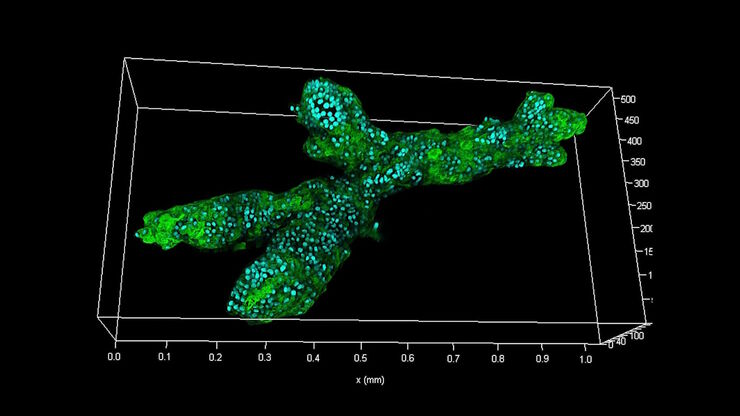
Examining Developmental Processes In Cancer Organoids
Interview: Prof. Bausch and Dr. Pastucha, Technical University of Munich, discuss using microscopy to study development of organoids, stem cells, and other relevant disease models for biomedical…
Loading...
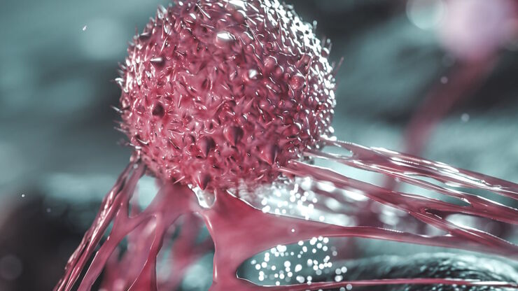
The Role of Iron Metabolism in Cancer Progression
Iron metabolism plays a role in cancer development and progression, and modulates the immune response. Understanding how iron influences cancer and the immune system can aid the development of new…
Loading...
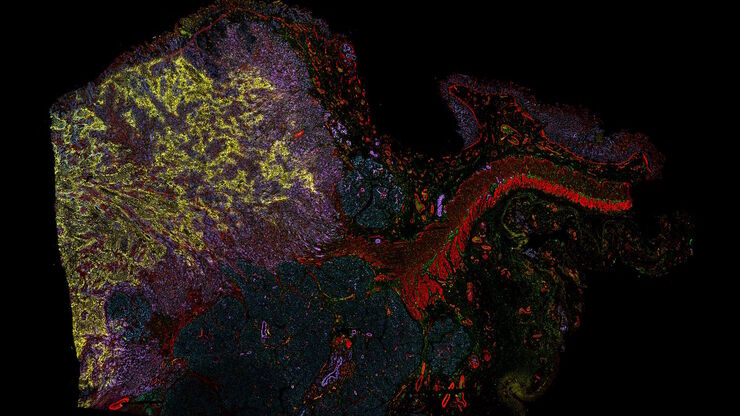
Dig Deeper Into the Complexities of Pancreatic Cancer with Multiplex Imaging
Cell DIVE is an iterative staining workflow for multiplexed imaging that unveils biological pathways to dig deeper into the complexities of pancreatic cancer.
Loading...
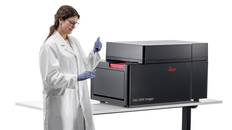
Complex Made Simple: Antibodies in Multiplexed Imaging
Build panels, plan studies, and get the most from precious reagents using this antibody multiplexing guide from Leica Microsystems
Loading...
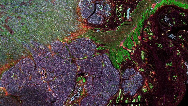
Multiplexed Imaging Types, Benefits and Applications
Multiplexed imaging is an emerging and exciting way to extract information from human tissue samples by visualizing many more biomarkers than traditional microscopy. By observing many biomarkers…
Loading...
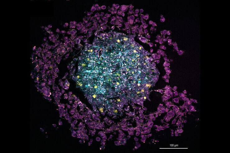
The Potential of Coherent Raman Scattering Microscopy at a Glance
Coherent Raman scattering microscopy (CRS) is a powerful approach for label-free, chemically specific imaging. It is based on the characteristic intrinsic vibrational contrast of molecules in the…
Loading...

Imaging of Anti-Cancer Drug Uptake in Spheroids using DLS
Spheroid 3D cell culture models mimic the physiology and functions of living tissues making them a useful tool to study tumor morphology and screen anti-cancer drugs. The drug AZD2014 is a recognized…
Loading...

Be Confident in your Results with Cell DIVE Validated Antibodies
The Cell DIVE System includes a carefully curated list of hundreds of commercially available antibodies validated to offer optimal specificity and sensitivity in multiplexed imaging. That validation…


