
Science Lab
Science Lab
The knowledge portal of Leica Microsystems offers scientific research and teaching material on the subjects of microscopy. The content is designed to support beginners, experienced practitioners and scientists alike in their everyday work and experiments. Explore interactive tutorials and application notes, discover the basics of microscopy as well as high-end technologies – become part of the Science Lab community and share your expertise!
Filter articles
Tags
Story Type
Products
Loading...

Total Internal Reflection Fluorescence (TIRF) Microscopy
Total internal reflection fluorescence (TIRF) is a special technique in fluorescence microscopy developed by Daniel Axelrod at the University of Michigan, Ann Arbor in the early 1980s. TIRF microscopy…
Loading...

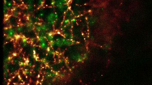
Applications of TIRF Microscopy in Life Science Research
The special feature of TIRF microscopy is the employment of an evanescent field for fluorophore excitation. Unlike standard widefield fluorescence illumination procedures with arc lamps, LEDs or…
Loading...
Super-Resolution GSDIM Microscopy
The nanoscopic technique GSDIM (ground state depletion microscopy followed by individual molecule return) provides a detailed image of the spatial arrangement of proteins and other biomolecules within…
Loading...

Optical Contrast Methods
Optical contrast methods give the potential to easily examine living and colorless specimens. Different microscopic techniques aim to change phase shifts caused by the interaction of light with the…
Loading...

Integrated Modulation Contrast (IMC)
Hoffman modulation contrast has established itself as a standard for the observation of unstained, low-contrast biological specimens. The integration of the modulator in the beam path of themodern…
Loading...

Polarization Contrast
Polarization microscopy is routinely applied in material sciences and geology to identify minerals on the basis of characteristic refraction properties and colors. In biology, polarization microscopy…
Loading...

Fluorescence in Microscopy
Fluorescence microscopy is a special form of light microscopy. It uses the ability of fluorochromes to emit light after being excited with light of a certain wavelength. Proteins of interest can be…
Loading...

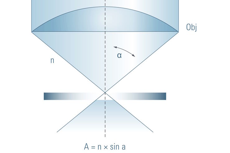
Depth of Field in Microscopy
In microscopy, depth of field is often seen as an empirical parameter. In practice it is determined by the correlation between numerical aperture, resolution and magnification. For the best possible…
Loading...
Beware of "Empty" Magnification
This article explains how to avoid the phenomen of "empty magnification" in microscopy.