
Science Lab
Science Lab
The knowledge portal of Leica Microsystems offers scientific research and teaching material on the subjects of microscopy. The content is designed to support beginners, experienced practitioners and scientists alike in their everyday work and experiments. Explore interactive tutorials and application notes, discover the basics of microscopy as well as high-end technologies – become part of the Science Lab community and share your expertise!
Filter articles
Tags
Products
Loading...

Rapidly Visualizing Magnetic Domains in Steel with Kerr Microscopy
The rotation of polarized light after interaction with magnetic domains in a material, known as the Kerr effect, enables the investigation of magnetized samples with Kerr microscopy. It allows rapid…
Loading...
 tissue.")
AI-Powered Multiplexed Image Analysis to Explore Colon Adenocarcinoma
In this application note, we demonstrate a spatial biology workflow via an AI-powered multiplexed image analysis-based exploration of the tumor immune microenvironment in colon adenocarcinoma.
Loading...

Tympanoplasty Surgery: Optimal Approaches and Tools
Discover tympanoplasty surgery case studies illustrating the standard approaches: post-auricular, endaural and transcanal. Gain insights from Dr. Flanagan on selecting the adequate surgical approach…
Loading...
.")
Dual-View LightSheet Microscope for Large Multicellular Systems
Visualizing the dynamics of complex multicellular systems is a fundamental goal in biology. To address the challenges of live imaging over large spatiotemporal scales, Franziska Moos et. al. present…
Loading...

A Meta-cancer Analysis of the Tumor Spatial Microenvironment
Learn how clustering analysis of Cell DIVE datasets in Aivia can be used to understand tissue-specific and pan-cancer mechanisms of cancer progression
Loading...
 tissue.")
Mapping the Landscape of Colorectal Adenocarcinoma with Imaging and AI
Discover deep insights in colon adenocarcinoma and other immuno-oncology realms through the potent combination of multiplexed imaging of Cell DIVE and Aivia AI-based image analysis
Loading...

Spatial Architecture of Tumor and Immune Cells in Tumor Tissues
Dig deep into the spatial biology of cancer progression and mouse immune-oncology in this poster, and learn how tumor metabolism can effect immune cell function.
Loading...

IBEX, Cell DIVE, and RNA-Seq: A Multi-omics Approach to Follicular Lymphoma
In a recent study by Radtke et al., a multi-omics spatial biology approach helps shed light on early relapsing lymphoma patients
Loading...
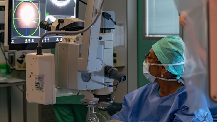
RPE65 Gene Therapy Subretinal Injection: Benefits of Intraoperative OCT
Discover how RPE65 gene therapy subretinal injection procedures in patients with Leber congenital amaurosis is supported by intraoperative Optical Coherence Tomography.