
Science Lab
Science Lab
The knowledge portal of Leica Microsystems offers scientific research and teaching material on the subjects of microscopy. The content is designed to support beginners, experienced practitioners and scientists alike in their everyday work and experiments. Explore interactive tutorials and application notes, discover the basics of microscopy as well as high-end technologies – become part of the Science Lab community and share your expertise!
Filter articles
Tags
Story Type
Products
Loading...
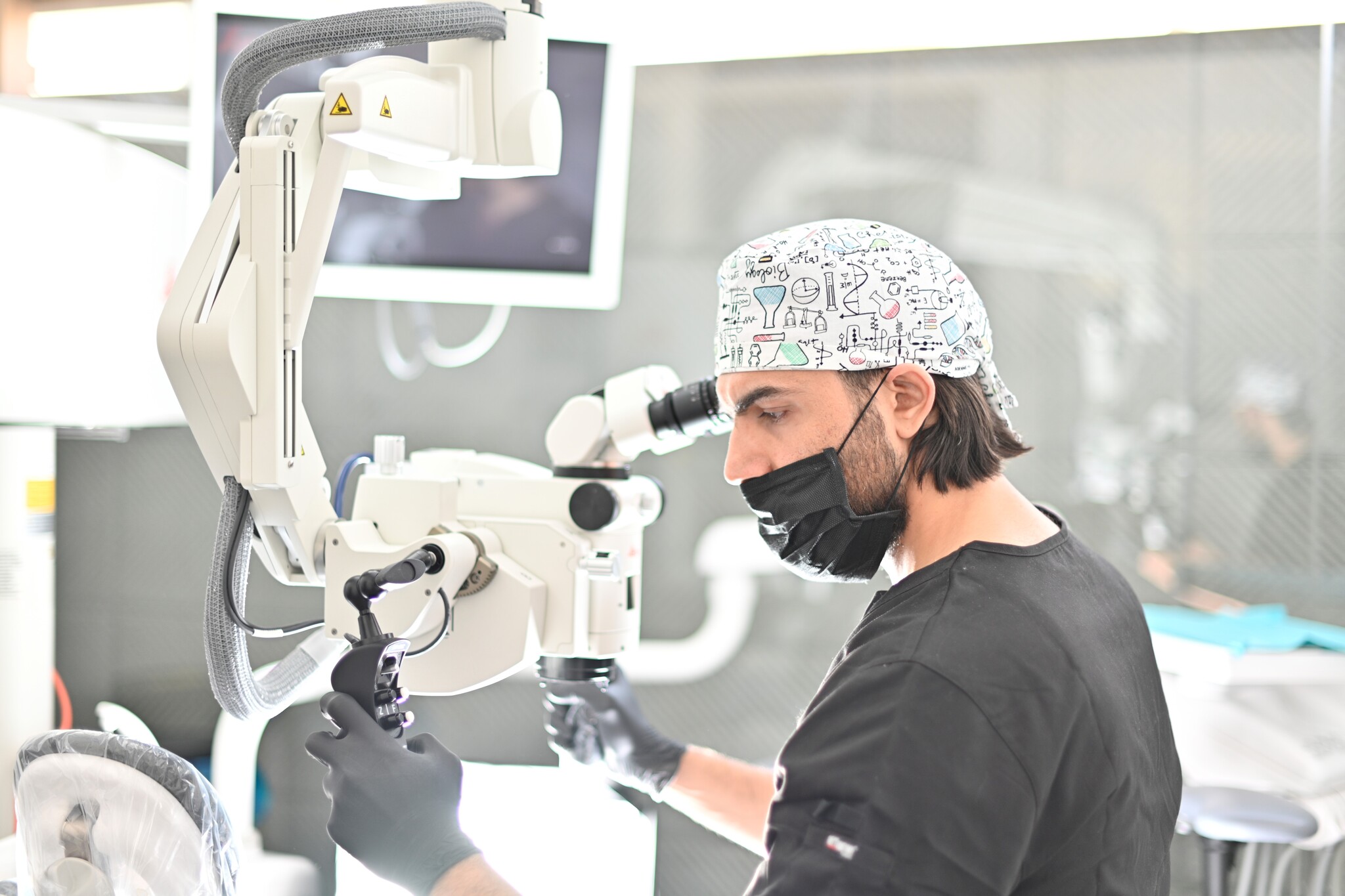
Minimally Invasive Dentistry: Visualization & Posture
Microscopes do not only provide better visualization during dental surgery. They also help ensure correct posture to avoid back pain and neck injuries. Dr. Iyad Ghoneim, from the Safad Dental Center…
Loading...
THUNDER Imagers: High Performance, Versatility and Ease-of-Use for your Everyday Imaging Workflows
This webinar will showcase the versatility and performance of THUNDER Imagers in many different life science applications: from counting nuclei in retina sections and RNA molecules in cancer tissue…
Loading...
 from the desired fluorescent signal from the cell membrane (green).")
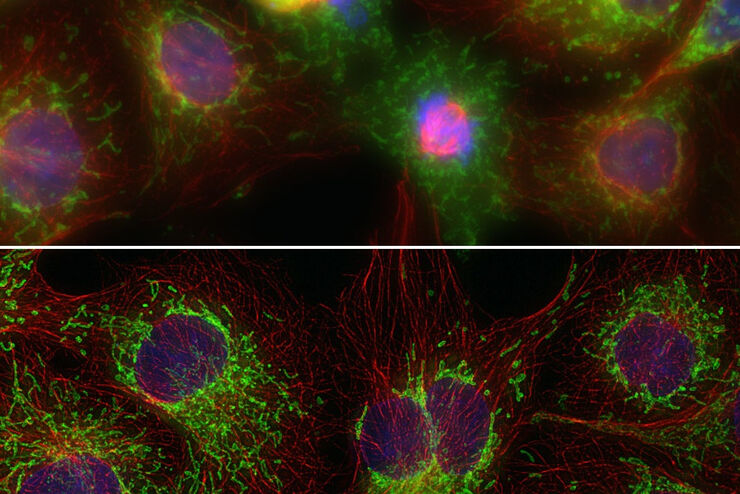
Learn how to Remove Autofluorescence from your Confocal Images
Autofluorescence can significantly reduce what you can see in a confocal experiment. This article explores causes of autofluorescence as well as different ways to remove it, from simple media fixes to…
Loading...
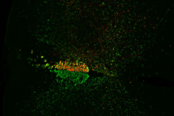
Evaluating Axon Regeneration After Brain or Spine Trauma of Mice
Damaged nerve regeneration was investigated using mouse spinal cord sections treated with compounds that counter axon growth inhibitor (AGI) proteins. The sections were screened to find active and…
Loading...
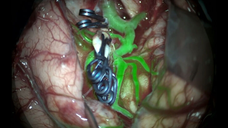
GLOW800 Augmented Reality Fluorescence in Aneurysm Treatment
This case study from Prof. Dr. Feres Chaddad talks about the treatment of unruptured MCA (middle cerebral artery) and PCOM (posterior communicating artery) aneurysms with microsurgical clipping. It…
Loading...
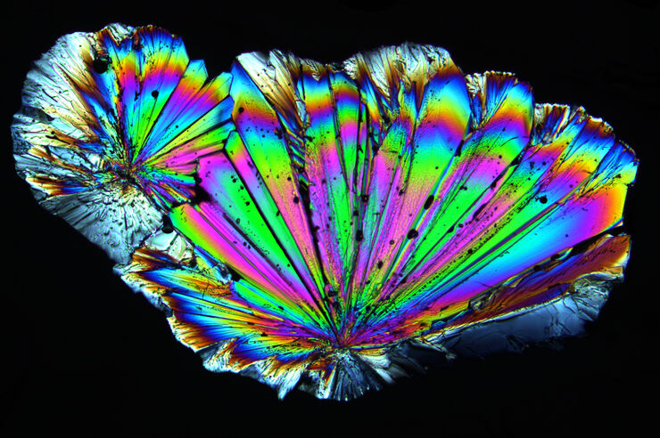
Polarizing Microscope Image Gallery
Polarized light microscopy (also known as polarizing microscopy) is an important method used in different fields, including research and quality assurance. It goes beyond just producing images at high…
Loading...
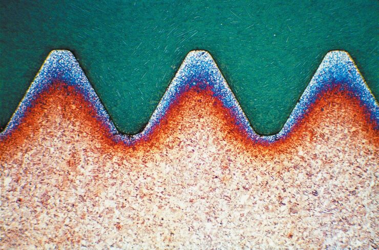
Metallography – an Introduction
This article gives an overview of metallography and metallic alloy characterization. Different microscopy techniques are used to study the alloy microstructure, i.e., microscale structure of grains,…
Loading...
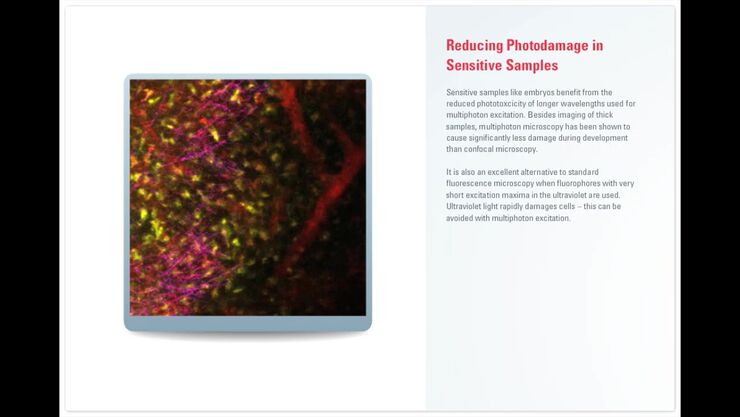
Principles of Multiphoton Microscopy for Deep Tissue Imaging
This tutorial explains the principles of multiphoton microscopy for deep tissue imaging. Multiphoton microscopy uses excitation wavelengths in the infrared taking advantage of the reduced scattering…
Loading...
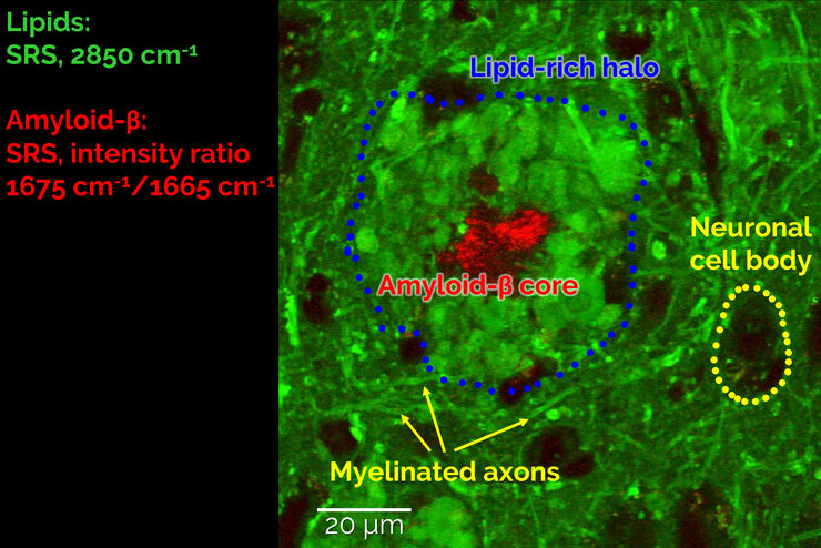
Stimulated Raman Scattering Microscopy Probes Neurodegenerative Disease
Despite decades of research, the molecular mechanisms underlying some of the most severe neurodegenerative diseases, such as Alzheimer’s or Parkinson’s, remain poorly understood. The progression of…