
Science Lab
Science Lab
The knowledge portal of Leica Microsystems offers scientific research and teaching material on the subjects of microscopy. The content is designed to support beginners, experienced practitioners and scientists alike in their everyday work and experiments. Explore interactive tutorials and application notes, discover the basics of microscopy as well as high-end technologies – become part of the Science Lab community and share your expertise!
Filter articles
Tags
Story Type
Products
Loading...
What is a Field-of-View Scanner?
A field-of-view scanner is an assembly of galvanometric scanning mirrors used in single-point confocal microscopes that offer the correct optical recording of large field sizes. The field-of-view…
Loading...

Resolved Field Number (RFN)
The field number (FN) for optical microscopes indicates the field of view (FOV). It corresponds to the area in the intermediate image that is observable through the eyepieces. Although, we cannot…
Loading...
Real Time Images of 3D Specimens with Sharp Contrast Free of Haze
THUNDER Imagers deliver in real time images of 3D specimens with sharp contrast, free of the haze or out-of-focus blur typical of widefield systems. They can even image clearly places deep inside a…
Loading...
How to improve your Alzheimer Protein Analysis with Laser Microdissection
Brain Research: Collect pure starting material for proteomics - Improve your workflow with Laser Microdissection - Many brain diseases result from protein malfunction, misfolding and agglutination.…
Loading...

What is a Tandem Scanner?
A Tandem Scanner is an assembly of two different types of scanning together in one system for true confocal point scanning. The Tandem Scanner consists of a three-mirror scanning base with the…
Loading...
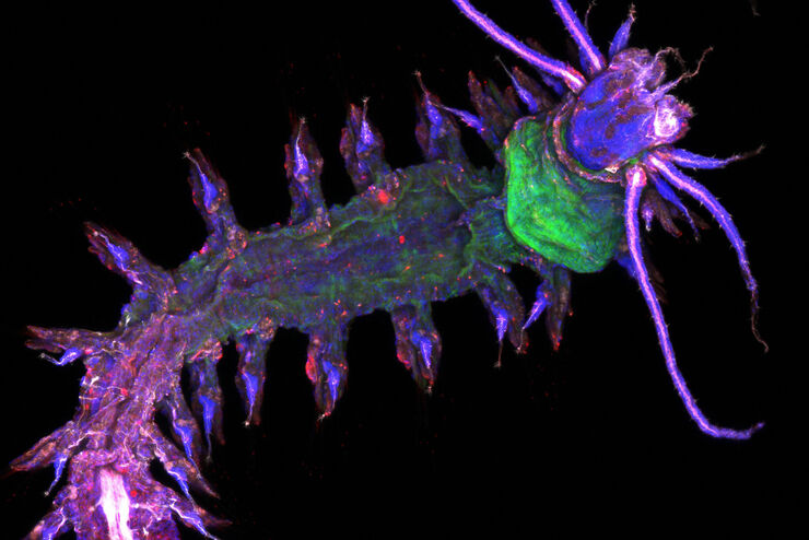
![3D glomeruli in a portion of an ECi-cleared kidney scanned by light sheet microscopy. Courtesy of Prof. Norbert Gretz, Medical Faculty Mannheim, University of Heidelberg [1].](/fileadmin/_processed_/d/d/csm_DLS-Sample-Preparation-Intr_527af241c5.jpg "3D glomeruli in a portion of an ECi-cleared kidney scanned by light sheet microscopy. Courtesy of Prof. Norbert Gretz, Medical Faculty Mannheim, University of Heidelberg [1].")
Using Mounting Frames for Light Sheet Sample Preparation
Sample handling is an important topic in the context of Light Sheet Microscopy. The TCS SP8 DLS integrates Light Sheet technology into an inverted confocal platform and can hence make use of general…
Loading...

How to improve your DNA Mutation Analysis Workflow with Laser Microdissection
DNA mutations lead to abnormal proteins or missing functional proteins, which can cause cells to multiply uncontrollably and become cancerous. To find and understand the underlying mutation for a…
Loading...

Using a Rotation Device for Light Sheet Sample Mounting
The TCS SP8 DLS from Leica Microsystems is an innovative concept to integrate the Light Sheet Microscopy technology into the confocal microscope. Due to its unique optical architecture samples can be…
Loading...
Array Tomography for SEM 3D Reconstruction
Array tomography (AT) is a 3D image reconstruction technique for high resolution, quantitative analysis of biological structures. For optimal results, ultrathin and ordered sections are an absolute…