
Science Lab
Science Lab
The knowledge portal of Leica Microsystems offers scientific research and teaching material on the subjects of microscopy. The content is designed to support beginners, experienced practitioners and scientists alike in their everyday work and experiments. Explore interactive tutorials and application notes, discover the basics of microscopy as well as high-end technologies – become part of the Science Lab community and share your expertise!
Filter articles
Tags
Story Type
Products
Loading...

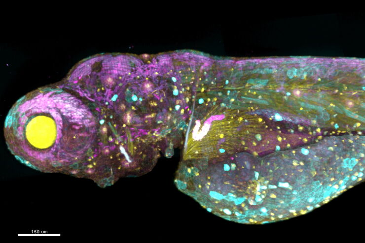
Multiplexing through Spectral Separation of 11 Colors
Fluorescence microscopy is a fundamental tool for life science research that has evolved and matured together with the development of multicolor labeling strategies in cells tissues and model…
Loading...

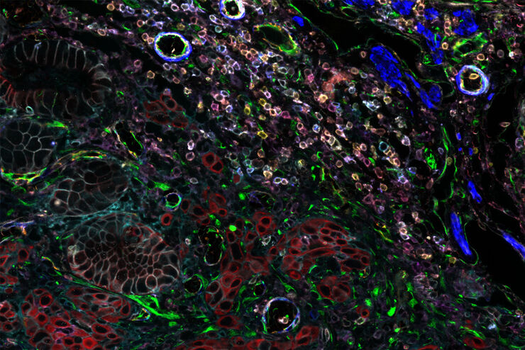
Multiplexed Imaging Types, Benefits and Applications
Multiplexed imaging is an emerging and exciting way to extract information from human tissue samples by visualizing many more biomarkers than traditional microscopy. By observing many biomarkers…
Loading...
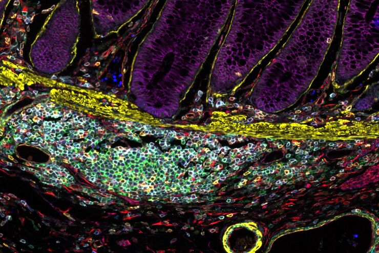
, Astrocytes (GFAP, red), Nuclei (DAPI, blue).")
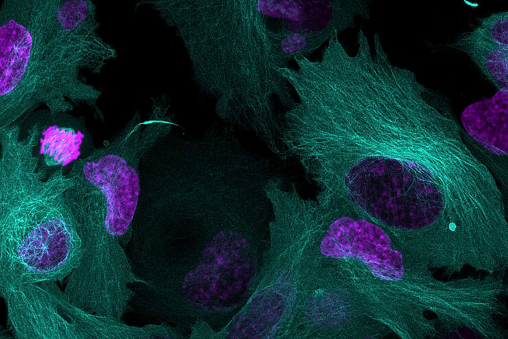
Multicolor Microscopy: The Importance of Multiplexing
The term multiplexing refers to the use of multiple fluorescent dyes to examine various elements within a sample. Multiplexing allows related components and processes to be observed in parallel,…
Loading...
A New Method for Convenient and Efficient Multicolor Imaging
The technique combining hyperspectral unmixing and phasor analysis was developed to simplify the process of getting images from a sample labeled with multiple fluorophores. This aggregate method…
Loading...
Considerations for Multiplex Live Cell Imaging
Simultaneous multicolor imaging for successful experiments: Live-cell imaging experiments are key to understand dynamic processes. They allow us to visually record cells in their living state, without…
Loading...
Designing your Research Study with Multiplexed IF Imaging
Multiplexed tissue analysis is a powerful technique that allows comparisons of cell-type locations and cell-type interactions within a single fixed tissue sample. It is common for researchers to ask…
Loading...
Be Confident in your Results with Cell DIVE Validated Antibodies
The Cell DIVE System includes a carefully curated list of hundreds of commercially available antibodies validated to offer optimal specificity and sensitivity in multiplexed imaging. That validation…