
Science Lab
Science Lab
The knowledge portal of Leica Microsystems offers scientific research and teaching material on the subjects of microscopy. The content is designed to support beginners, experienced practitioners and scientists alike in their everyday work and experiments. Explore interactive tutorials and application notes, discover the basics of microscopy as well as high-end technologies – become part of the Science Lab community and share your expertise!
Filter articles
Tags
Story Type
Products
Loading...
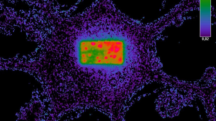
Förster Resonance Energy Transfer (FRET)
The Förster Resonance Energy Transfer (FRET) phenomenon offers techniques that allow studies of interactions in dimensions below the optical resolution limit. FRET describes the transfer of the energy…
Loading...
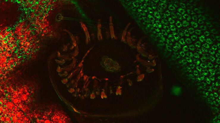
An Introduction to CARS Microscopy
CARS overcomes the drawbacks of conventional staining methods by the intrinsic characteristics of the method. CARS does not require labeling because it is highly specific to molecular compounds which…
Loading...
")
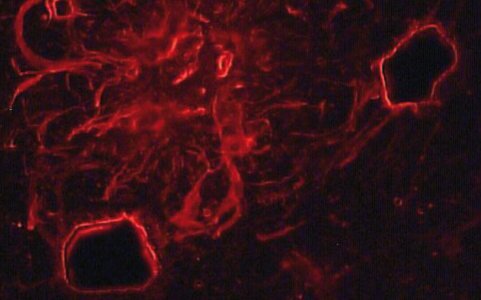
Mosaic Images
Confocal laser scanning microscopes are widely used to create highly resolved 3D images of cells, subcellular structures and even single molecules. Still, an increasing number of scientists are…
Loading...

An Introduction to Laser Microdissection
Laser microdissection is a highly selective process for preparing samples for DNA, RNA and protein analysis. It is a microscope-controlled manipulation technique for the precise separation of samples…
Loading...
Applications of Laser Microdissection
Laser microdissection and laser micromanipulation are suitable for gaining a differentiated insight into the function of genes and proteins, and are used for a wide range of applications in…
Loading...
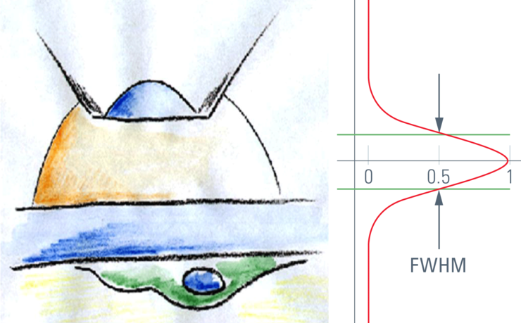
Confocal Optical Section Thickness
Confocal microscopes are employed to optically slice comparably thick samples.
Loading...

Optical Contrast Methods
Optical contrast methods give the potential to easily examine living and colorless specimens. Different microscopic techniques aim to change phase shifts caused by the interaction of light with the…
Loading...

Integrated Modulation Contrast (IMC)
Hoffman modulation contrast has established itself as a standard for the observation of unstained, low-contrast biological specimens. The integration of the modulator in the beam path of themodern…
Loading...

Polarization Contrast
Polarization microscopy is routinely applied in material sciences and geology to identify minerals on the basis of characteristic refraction properties and colors. In biology, polarization microscopy…