
Science Lab
Science Lab
The knowledge portal of Leica Microsystems offers scientific research and teaching material on the subjects of microscopy. The content is designed to support beginners, experienced practitioners and scientists alike in their everyday work and experiments. Explore interactive tutorials and application notes, discover the basics of microscopy as well as high-end technologies – become part of the Science Lab community and share your expertise!
Filter articles
Tags
Story Type
Products
Loading...

Basics in Component Cleanliness Analysis
An overview on the basics of component cleanliness and analysis solutions that can be tailored to your specific needs is presented. For the automotive industry, obtaining results rapidly, accurately,…
Loading...

Multiplexing through Spectral Separation of 11 Colors
Fluorescence microscopy is a fundamental tool for life science research that has evolved and matured together with the development of multicolor labeling strategies in cells tissues and model…
Loading...
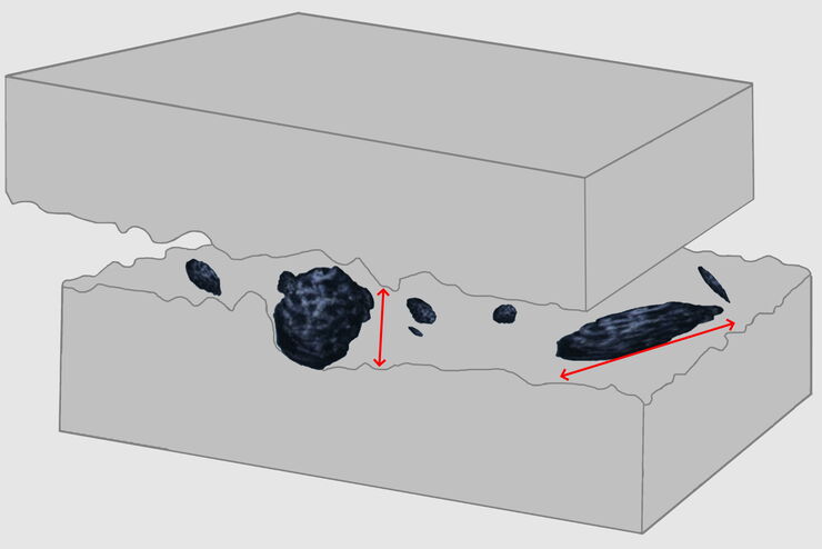
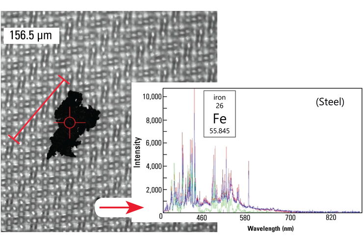
3 Factors Determine the Damage Potential of Particles
This article discusses the 3 factors for determining the potential of a particle to cause damage to parts and components in the automotive and electronic industry. These factors include the…
Loading...

Skull Base Neurosurgery: Epidural Lateral Approaches
Surgery of skull base tumors and diseases, such as cavernomas, epidermoid cysts, meningiomas and schwannomas, can be quite complex. During the Leica 2021 Neurovisualization Summit, a unique event…
Loading...

Studying Ocular Birth Defects
This article discusses how lens formation and ocular birth defects can be studied with sharp widefield microscopy images which are acquired rapidly. The mouse ocular lens is used as a model to study…
Loading...
 and CellEvent™ (yellow).")
Following Multiple Events during Staurosporine Apoptosis
Coming next on MicaCam - Livestream on 19th October 2022 - In this episode of MicaCam, we show how adding additional markers to an apoptosis kit can markedly increase the amount of information a…
Loading...
Cleanliness Analysis with a 2-Methods-in-1 Solution
In this article, it is examined how an overall efficient and cost-effective cleanliness analysis workflow can be achieved with a 2-methods-in-1 materials analysis solution, combining optical…
Loading...

Precise Spatial Proteomic Information in Tissues
Despite the availability of imaging-based and mass-spectrometry-based methods for spatial proteomics, a key challenge remains connecting images with single-cell-resolution protein abundance…
Loading...

RNA Quality after Different Tissue Sample Preparation
The influence of sample preparation and ultraviolet (UV) laser microdissection (UV LMD) on the quality of RNA from murine-brain tissue cryo-sections is described in this article. To obtain good…