
Science Lab
Science Lab
The knowledge portal of Leica Microsystems offers scientific research and teaching material on the subjects of microscopy. The content is designed to support beginners, experienced practitioners and scientists alike in their everyday work and experiments. Explore interactive tutorials and application notes, discover the basics of microscopy as well as high-end technologies – become part of the Science Lab community and share your expertise!
Filter articles
Tags
Story Type
Products
Loading...

Digitalization in Neurosurgical Planning and Procedures
Learn about Augmented Reality, Virtual Reality and Mixed Reality in neurosurgery and how they can help overcome challenges.
Loading...

Augmented Reality Assisted Navigation in Neuro-Oncological Surgery
In neuro-oncological surgery, new technologies such as Augmented Reality are helping to improve surgical precision enabling a precise trajectory, conformational resection, the absence of collateral…
Loading...
Factors to Consider for a Cleanliness Analysis Solution
Choosing the right cleanliness analysis solution is important for optimal quality control. This article discusses the important factors that should be taken into account to find the solution that best…
Loading...

New Imaging Tools for Cryo-Light Microscopy
New cryo-light microscopy techniques like LIGHTNING and TauSense fluorescence lifetime-based tools reveal structures for cryo-electron microscopy.
Loading...

H&E Staining in Microscopy
If we consider the role of microscopy in pathologists’ daily routines, we often think of the diagnosis. While microscopes indeed play a crucial role at this stage of the pathology lab workflow, they…
Loading...
, the cis-golgi matrix protein GM130 (AF488, green), and the trans-golgi network membrane protein TGN46 (AF647, red).")
Golgi Organizational Changes in Response to Cell Stress
VIDEO ON DEMAND - In this episode of MicaCam, our special guest George Galea from EMBL Heidelberg will look at HeLa Kyoto cells treated with various chemotherapeutic agents to investigate their effect…
Loading...

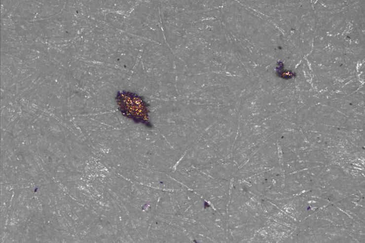
Cleanliness Analysis for Particulate Contamination
Devices, products, and their components fabricated in many industries can be quite sensitive to contamination and, as a result, have stringent requirements for technical cleanliness. Measurement…
Loading...

Efficient Particle Counting and Analysis
This report discusses particle counting and analysis using optical microscopy for cleanliness of parts and components. Particle counting and analysis is a critical part of quality assurance in the…
Loading...
 stained to show the nucleus")
AI-Enabled Spatial Analysis of Complex 3D Datasets
VIDEO ON DEMAND - This edition of MicaCam offers practical advice on the extraction of publication grade insights from microscopy images. Our special guest Luciano Lucas (Leica Microsystems) will…