
Science Lab
Science Lab
The knowledge portal of Leica Microsystems offers scientific research and teaching material on the subjects of microscopy. The content is designed to support beginners, experienced practitioners and scientists alike in their everyday work and experiments. Explore interactive tutorials and application notes, discover the basics of microscopy as well as high-end technologies – become part of the Science Lab community and share your expertise!
Filter articles
Tags
Story Type
Products
Loading...

Benefits of Fluorescence in Vascular Neurosurgery
Fluorescein and ICG fluorescence videoangiography have transformed the experience of vascular neurosurgeons, providing an intraoperative view with enriched information. During the Leica 2021…
Loading...

Quality Control Under the Microscope
Fast-rising demand for electric vehicles is one of the market’s main drivers, but there are other hotspots of growth, including the rise in renewable energy installations, such as photovoltaic panels,…
Loading...

Multiplexed Imaging Types, Benefits and Applications
Multiplexed imaging is an emerging and exciting way to extract information from human tissue samples by visualizing many more biomarkers than traditional microscopy. By observing many biomarkers…
Loading...

3D Tissue Imaging: From Fast Overview To High Resolution With One Click
3D Tissue imaging is a widespread discipline in the life sciences. Researchers use it to reveal detailed information of tissue composition and integrity, to make conclusions from experimental…
Loading...

How To Perform Fast & Stable Multicolor Live-Cell Imaging
With the help of live-cell imaging researchers gain insights into dynamic processes of living cells up to whole organisms. This includes intracellular as well as intercellular activities. Protein or…
Loading...

Imaging of Cardiac Tissue Regeneration in Zebrafish
Learn how to image cardiac tissue regeneration in zebrafish focusing on cell proliferation and response during recovery. MicaCam Episode 04 with Laura Peces-Barba Castaño from the Max Planck…
Loading...

Multiplexed Imaging Glossary
Human tissues contain a many different biomarkers which can be explored to reveal the workings of biological pathways and interrogate disease states. Recent technological advances have made it…
Loading...

Hydraulics in the Automotive and Aerospace Industries
Cleanliness standards relating to lubricants, hydraulic fluids, and oils, e.g., ISO 4406 and DIN 51455 are discussed in this article. Cleanliness plays a central role in the automotive and…
Loading...
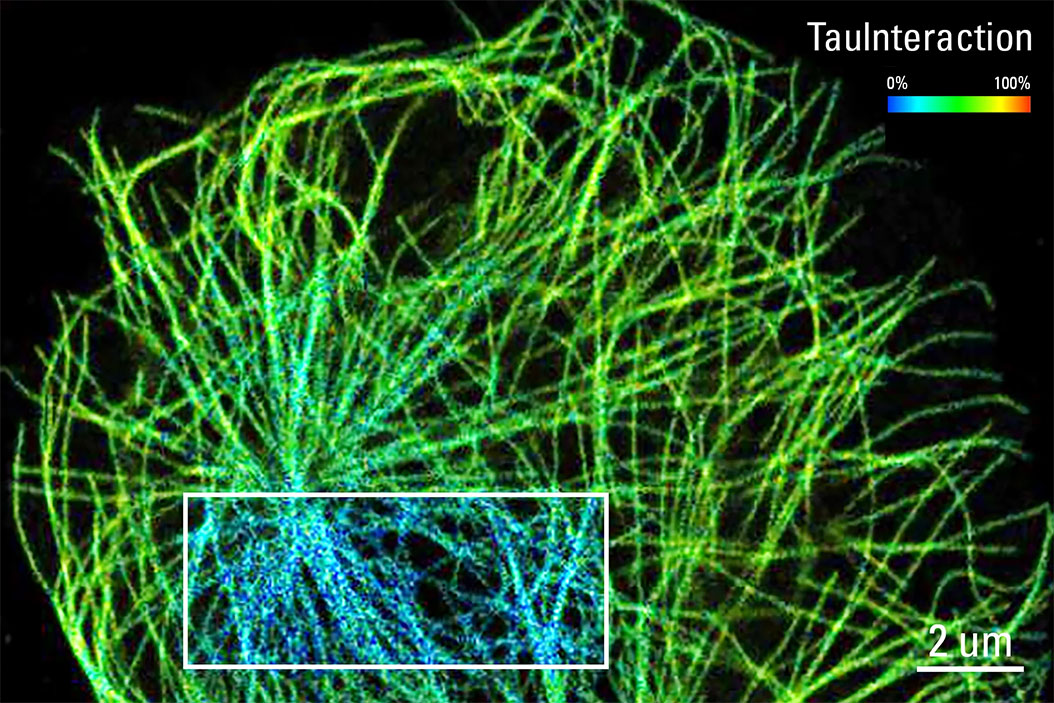
TauInteraction – Studying Molecular Interactions with TauSense
Fluorescence microscopy constitutes one of the pillars in life sciences and is a tool commonly used to unveil cellular structure and function. A key advantage of fluorescence microscopy resides in the…