
Science Lab
Science Lab
The knowledge portal of Leica Microsystems offers scientific research and teaching material on the subjects of microscopy. The content is designed to support beginners, experienced practitioners and scientists alike in their everyday work and experiments. Explore interactive tutorials and application notes, discover the basics of microscopy as well as high-end technologies – become part of the Science Lab community and share your expertise!
Filter articles
Tags
Story Type
Products
Loading...

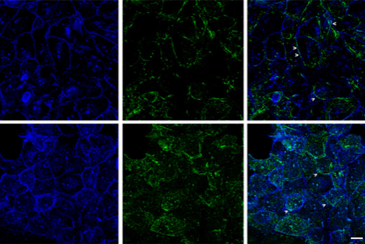
Physiology Image Gallery
Physiology is about the processes and functions within a living organism. Research in physiology focuses on the activities and functions of an organism’s organs, tissues, or cells, including the…
Loading...
 and astrocytes (green) in a cortical spheroid derived from human induced pluripotent stem cells.")
Neuroscience Images
Neuroscience commonly uses microscopy to study the nervous system’s function and understand neurodegenerative diseases.
Loading...

Developmental Biology Image Gallery
Developmental biology explores the development of complex organisms from the embryo to adulthood to understand in detail the origins of disease. This category of the gallery shows images about…
Loading...
, specific neuronal markers (magenta) and cell nuclei (white), computational cleared.")
Into the Third Dimension with "Wow Effect"- Observe Cells in 3D and Real-Time
Life is fast, especially for a cell. As a rule, cells should be examined under physiological conditions which are as close as possible to their natural environment. New technologies offer tremendous…
Loading...
 and astrocytes (green) in a cortical spheroid derived from human induced pluripotent stem cells.")
Download The Guide to Live Cell Imaging
In life science research, live cell imaging is an indispensable tool to visualize cells in a state as in vivo as possible. This E-book reviews a wide range of important considerations to take to…
Loading...

Targeting Active Recycling Nuclear Pore Complexes using Cryo Confocal Microscopy
In this article, how cryo light microscopy and, in particular cryo confocal microscopy, is used to improve the reliability of cryo EM workflows is described. The quality of the EM grids and samples is…
Loading...
 and cytokinesis ring (Rlc1-mCherry; red).")
Studying Cell Division
Cell division is a biological process during which all cellular components must be distributed among the daughter cells. The division process requires firm coordination for success. Microscopy is…
Loading...

The Power of Pairing Adaptive Deconvolution with Computational Clearing
Learn how deconvolution allows you to overcome losses in image resolution and contrast in widefield fluorescence microscopy due to the wave nature of light and the diffraction of light by optical…
Loading...
Improvement of Imaging Techniques to Understand Organelle Membrane Cell Dynamics
Understanding cell functions in normal and tumorous tissue is a key factor in advancing research of potential treatment strategies and understanding why some treatments might fail. Single-cell…