
Science Lab
Science Lab
The knowledge portal of Leica Microsystems offers scientific research and teaching material on the subjects of microscopy. The content is designed to support beginners, experienced practitioners and scientists alike in their everyday work and experiments. Explore interactive tutorials and application notes, discover the basics of microscopy as well as high-end technologies – become part of the Science Lab community and share your expertise!
Filter articles
Tags
Story Type
Products
Loading...
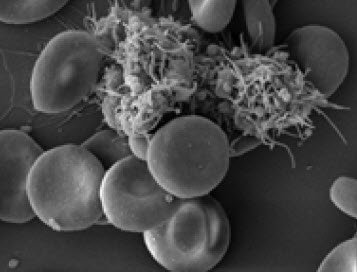
Human Blood Cells Protocol
Application Note for Leica EM CPD300 - Life Science Research. Species: Human (Homo sapiens)
Critical point drying of human blood with subsequent platinum / palladium coating and SEM analysis.
Loading...

Six Features to Consider when Choosing a Dental Microscope
In dental medicine, the surgical microscope has become increasingly important for high-quality and successful surgeries, particularly in the field of endodontics. A microscope supports the dentist to…
Loading...
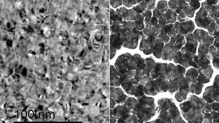
Improvement of Metallic Thin Films for HR-SEM by Using DC Magnetron Sputter Coater
Preparation techniques, like several kinds of coating methods play an important role for high resolution scanning electron microscopy (HR-SEM). Nonconductive sample like biological and synthetic…
Loading...

Infinity Optical Systems
“Infinity Optics” refers to the concept of a beam path with parallel rays between the objective and the tube lens of a microscope. Flat optical components can be brought into this “Infinity Space”…
Loading...
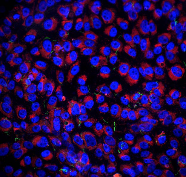
Imaging of Host Cell-bacteria Interactions using Correlative Microscopy under Cryo-conditions
Pathogenic bacteria have developed intriguing strategies to establish and promote infections in their respective hosts. Most bacterial pathogens initiate infectious diseases by adhering to host cells…
Loading...

Cataract Surgery with CoAx4 Illumination
A stable red reflex is one of the most important features of an ophthalmic surgical microscope for cataract surgery. It’s the red reflex that makes the structure of the lens visible and thus makes for…
Loading...
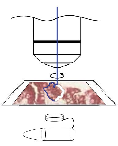
Workflows & Protocols: How to Isolate Individual Chromosomes with Laser Microdissection
During the first Leica Workshop in Brazil, at the Centro de Energia Nuclear na Agricultura/USP (CENA), the participants learned how to prepare samples for laser microdissection (LMD) using a cryotome.…
Loading...
")
A Brief History of Light Microscopy
The history of microscopy begins in the Middle Ages. As far back as the 11th century, plano-convex lenses made of polished beryl were used in the Arab world as reading stones to magnify manuscripts.…
Loading...

From Light to Mind: Sensors and Measuring Techniques in Confocal Microscopy
This article outlines the most important sensors used in confocal microscopy. By confocal microscopy, we mean "True Confocal Scanning", i.e. the technique that illuminates and measures one single…