
Science Lab
Science Lab
The knowledge portal of Leica Microsystems offers scientific research and teaching material on the subjects of microscopy. The content is designed to support beginners, experienced practitioners and scientists alike in their everyday work and experiments. Explore interactive tutorials and application notes, discover the basics of microscopy as well as high-end technologies – become part of the Science Lab community and share your expertise!
Filter articles
Tags
Story Type
Products
Loading...
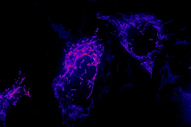
AI Microscopy Image Analysis – An Introduction
Artificial intelligence-guided microscopy image analysis and visualization is a powerful tool for data-driven scientific discovery. AI can help researchers tackle challenging imaging applications,…
Loading...
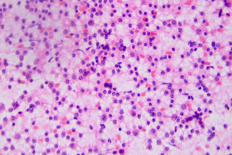
How to Benefit from Digital Cytopathology
If you have thought of digital cytopathology as characterized by the digitization of glass slides, this webinar with Dr. Alessandro Caputo from the University Hospital of Salerno, Italy will broaden…
Loading...
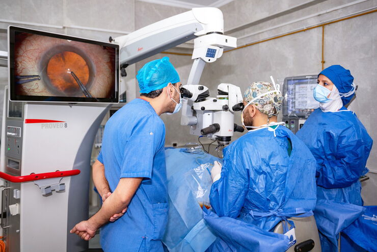
Dr. Tawfik Shares his Expert View on Direct Horizontal Chopping in Cataract Surgery
It is estimated that nearly 28 million cataract surgery procedures are performed worldwide every year. Phacoemulsification is the most common method used to remove the cataract and chopping maneuvers…
Loading...
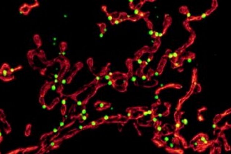
Multicolor 4D Super Resolution Light Sheet Microscopy
The AI Microscopy Symposium offers a unique forum for discussing the latest AI-based technologies and tools in the field of microscopy and biomedical imaging. In this scientific presentation, Yuxuan…
Loading...
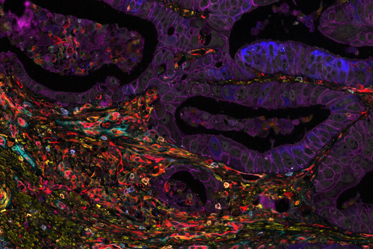
Hyperplex Cancer Tissue Analysis at Single Cell Level with Cell DIVE
The ability to study how lymphoma cell heterogeneity is influenced by the cells’ response to their microenvironment, especially at the mutational, transcriptomic, and protein levels. Protein…
Loading...
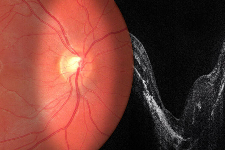
Clinical Symposium on OCT-Guided Retina Surgery
In this recording Prof. Tan from Singapore National Eye Centre and Dr. Català from Sant Joan de Déu Barcelona Children’s Hospital share their expertise on retinal surgery with intraoperative OCT from…
Loading...
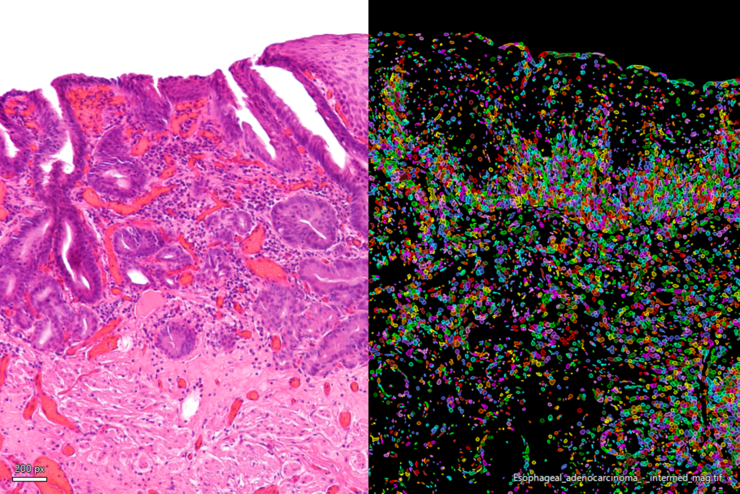
Simplifying the Cancer Biology Image Analysis Workflow
As cancer biology data sets grow, so do the challenges in microscopy image analysis. Aivia experts cover how to overcome these challenges with AI.
Loading...
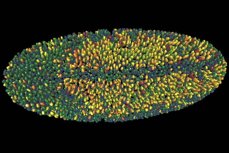
Examining Critical Developmental Events in High-Definition
Extended live cell imaging of embryo development requires a delicate balance between light exposure, temporal resolution and spatial resolution to maintain cells’ viability. Compromises between the…
Loading...
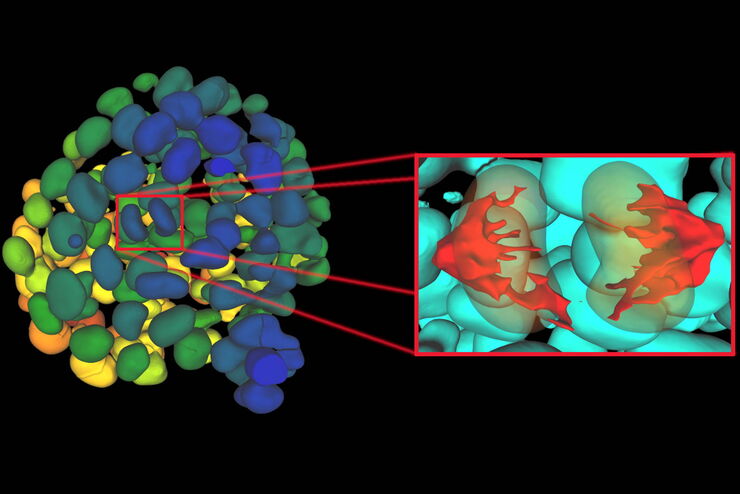
Observing Complex Cellular Interactions at Multiple Scales
Learn how to observe challenging cellular interactions with easy to deploy object detection and relationship measurements.