
Science Lab
Science Lab
The knowledge portal of Leica Microsystems offers scientific research and teaching material on the subjects of microscopy. The content is designed to support beginners, experienced practitioners and scientists alike in their everyday work and experiments. Explore interactive tutorials and application notes, discover the basics of microscopy as well as high-end technologies – become part of the Science Lab community and share your expertise!
Filter articles
Tags
Story Type
Products
Loading...
 and zinc (Zn).")
High Speed for Your Material Analysis Workflow
Learn from our expert, Dr. Konstantin Kartaschew, how the intelligent combination of light microscopy with laser-induced breakdown spectroscopy (LIBS) will truly accelerate your root-cause-analysis…
Loading...

How to Boost your Microelectronic Component Inspection Performance
Do you need to see more when inspecting silicon wafers or MEMS? Would you like to get sharp and detailed sample images which are similar to those from electron microscopes?
Watch this free webinar…
Loading...

How Industrial Applications Benefit from Fluorescence Microscopy
Watch this free webinar to know more about what you can do with fluorescence microscopy for industrial applications. We will cover a wide range of investigations where fluorescence contrast offers new…
Loading...

Intravital Microscopy of Cancer
Join our guest speaker Prof Dr Jacco van Rheenen, as he presents his work on the identity, behavior and fate of cells that drive the initiation and progression of cancer.
Loading...

Accelerating Neuron Image Analysis with Automation
The ability to examine complex neural processes relies on the accurate reconstruction of neuronal networks at scale. Most data extraction methods in neuroscience research are time-consuming and…
Loading...
Tracking Single Cells Using Deep Learning
AI-based solutions continue to gain ground in the field of microscopy. From automated object classification to virtual staining, machine and deep learning technologies are powering scientific…
Loading...

Learning the Cellular Architecture from its Optical Properties
In the last 3 years, microscopists have started to use "AI based" solutions for a wide range of applications, including image acquisition optimization (smart microscopy), object classification, image…
Loading...

How to Use a Digital Microscope to Streamline Inspection Processes
Watch this webinar for inspiration and expert advice on how to make quality control simpler, quicker, and easier. Learn how to perform comprehensive visual inspection, including comparison,…
Loading...
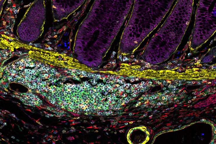
Designing your Research Study with Multiplexed IF Imaging
Multiplexed tissue analysis is a powerful technique that allows comparisons of cell-type locations and cell-type interactions within a single fixed tissue sample. It is common for researchers to ask…