
Science Lab
Science Lab
The knowledge portal of Leica Microsystems offers scientific research and teaching material on the subjects of microscopy. The content is designed to support beginners, experienced practitioners and scientists alike in their everyday work and experiments. Explore interactive tutorials and application notes, discover the basics of microscopy as well as high-end technologies – become part of the Science Lab community and share your expertise!
Filter articles
Tags
Story Type
Products
Loading...

Topographic Analysis of Firing Pin Impressions on Cartridge Cases
The analysis of fired cartridges for primer cup morphology and flattening and firing pin impression (crater) depth using topographic data is discussed in this article. Topographical analysis of the…
Loading...
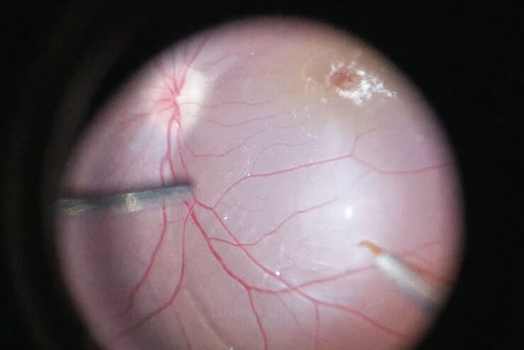
Intraoperative OCT-Assisted Gene Therapy
Gene augmentation therapy is a method of ocular gene transfer for autosomal recessive or X-linked retinal dystrophies when there is insufficient functional protein manifesting as genetic disease.…
Loading...

How does an Automated Rating Solution for Steel Inclusions Work?
The rating of non-metallic inclusions (NMIs) to determine steel quality is critical for many industrial applications. For an efficient and cost-effective steel quality evaluation, an automated NMI…
Loading...

An Introduction to Computational Clearing
Many software packages include background subtraction algorithms to enhance the contrast of features in the image by reducing background noise. The most common methods used to remove background noise…
Loading...

Challenges Faced When Manually Rating Non-Metallic Inclusions (NMIs) to Determine Steel Quality
Rapid, accurate, and reliable rating of non-metallic inclusions (NMIs) is instrumental for the determination of steel quality. This article describes the challenges that arise from manual NMI rating,…
Loading...

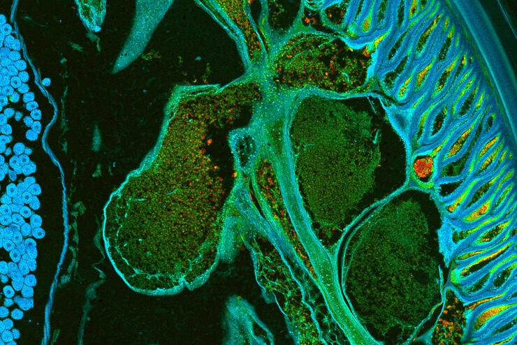
Expanding the Frontiers of Confocal Live Cell Imaging
Here we explore how STELLARIS unlocks the full power and potential of live cell studies by overcoming many common limitations and fully integrating fluorescence lifetime-based information to add a new…
Loading...
How to Uncover Hidden Dimensions in Research with Lifetime Imaging
Learn how fluorescence lifetime imaging adds information depth to your confocal experiments and reveals novel insights that are difficult or impossible to discover using conventional intensity-based…
Loading...
 from the desired fluorescent signal from the cell membrane (green).")
Learn how to Remove Autofluorescence from your Confocal Images
Autofluorescence can significantly reduce what you can see in a confocal experiment. This article explores causes of autofluorescence as well as different ways to remove it, from simple media fixes to…
Loading...
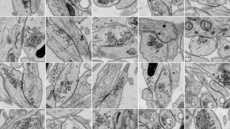
Bridging Structure and Dynamics at the Nanoscale through Optogenetics and Electrical Stimulation
Nanoscale ultrastructural information is typically obtained by means of static imaging of a fixed and processed specimen. However, this is only a snapshot of one moment within a dynamic system in…